Thursday, December 31, 2009

Microscope CCD Camera

The MW5CCD microscope camera has 5.6 mega pixels and includes software. The camera threads directly onto your microscope's c-mount adapter.

The included software can be used to make measurements, adjust color settings and capture and save both still and moving images.

Learn more about this microscope CCD camera here.

Tuesday, December 29, 2009

Microscope Boom Stands

Microscope boom stands allow the user to place large objects under the microscope. The stands provide ease of use, as they swing the microscope body toward and away from the items that need to be magnified.

The ball bearing boom stand slides smoothly along the horizontal axis. This horizontal axis can be secured on the vertical post in a number of varying heights.

Above is an example of an electronics inspection microscope system on a boom stand. This microscope system uses a 150w halogen dual arm fiber optic illuminator that provides pinpointed light for viewing printed circuit boards.

The articulated arm microscope stand has several joints that allow the microscope body to be placed in varying positions. This boom stand secures the microscope body in position until it is adjusted. It does not slide on ball bearings the same way that the ball bearing boom stand does.

Monday, December 28, 2009

Paulownia Wood Under Microscope

Paulownia wood is native to much of Asia. The tree grows extremely fast and has been used for reforestation, to make surfboards, and wood furniture.

Cross section of Paulownia wood captured at 100x magnification using a Nikon Coolpix 5400 camera adapter on a compound microscope.

Same cross-section captured at 200x magnification.

A variety of camera adapters for point and shoot and digital SLR cameras are available here.

Thursday, December 24, 2009

Measuring with Microscope Cameras

Making measurements with a microscope can be performed using an eyepiece reticle. Another way to perform measurements is by using the software that is included with a microscope camera. The image below was captured with the Lumenera Infinity 1-3 DK3000 3.1 mega pixel digital microscope camera, using the measuring software. The software was calibrated with the microscope and then measurements were made by using the computer.

Image captured at 1000x magnification using a metallurgical microscope.

When using the software to make measurements, the accuracy of the measurement is dependent on the ability to find the edges of what you are measuring. Measurements are made by drawing a line on the image. You can also draw a circle to determine the diameter or radius of the circle, or polygon images.

Tuesday, December 22, 2009

Magnifying Electronics

Printed circuit boards and small electronic parts can be difficult to view under a microscope because most of the components are not on a flat plane. These images were captured with a Meiji EMZ-5TR stereo microscope and a MC2000 digital camera at 15x magnification.

When viewing electronic parts, low magnification is generally best - anything between 5x - 20x is usually all that is required.

Adjusting the focus will allow you to view different focal depths in the undulating surfaces. Another option is to hold the piece in your hand and slowly move it up and down until the focus is crisp. Many technicians do this as they are soldering small pieces.

Monday, December 21, 2009

USB Microscopes

Digital microscopes, also known as USB microscopes allow you to connect the microscope directly to your computer. The microscope comes with software that when open, allows you to view a live image from the microscope directly on your computer.

USB microscopes allow you to gather multiple students around the computer to see an image, rather than taking turns to view a single image through the microscope eyepiece.

The software included with digital microscopes allows you to capture and save both still images and motion video. You can print images, email them, or put them into a presentation.

The Digital BA210 laboratory microscope is commonly found in Universities.

Digital USB microscopes are made for kids as well. This kids microscope is the DS-2 microscope. Low magnification of 20x and 40x is perfect for viewing rocks, coins, flowers or details on a toy car.

Friday, December 18, 2009

Brightfield, Darkfield and Phase Contrast

The images below are of a spleen and were captured at 400x magnification with the Meiji MT5310 microscope. This microscope has the capability of viewing specimens with brightfield, darkfield and phase contrast.

This image was captured using brightfield. Brightfield microscopy is the standard form of microscopy found in most high schools when using a high power light microscope.

This image was captured using darkfield. Darkfield is similar to back-lighting a subject. This microscopy technique takes the light and diffuses it toward the sides rather than directly through the objective lens.


This image was captured using phase contrast. Phase contrast is often used to view specimens that are the same color as the background they are against. Sometimes rather than staining a specimen, phase contrast will be used to produce similar results.

Wednesday, December 16, 2009

Actinopod Under Microscope

An actinopod is a protozoa with stiff rod-like pseudopods that radiate from the center. You can learn more about protozoans through these science videos and teacher handbooks.

Tuesday, December 15, 2009

Microscopic Worms

Check out these microscopic worms. Out of all known and studied worms, these have the DNA closest to human DNA.


Images were captured directly to the computer using the MC2300 3.0 mega pixel microscope camera on the SMZ-168 stereo zoom microscope with a 1.0 C-Mount Adapter. Images were captured at 100x magnification.

Monday, December 14, 2009

Low Power Microscopes

Low power or stereo microscopes are great for looking at things you might hold in your hand. These microscopes allow you to view details in objects that you can already see with the naked eye. Sometimes these microscopes will use no light, such as the 185 elementary microscope. Other stereo low power microscopes will have top and bottom illumination, such as the 460TBL microscope.

The amount of things you can view with a low power microscope is endless.

Paper clip at 20x magnification, captured with MC352 camera.

Penny at 7x magnification, captured with the MC352 camera.

Looking for other things to look at over the holiday with your kids? Check out the list below for some fun ideas of items you can view with your low power stereo microscope.
  • Dollar bill
  • Newspaper print
  • Twigs & leaves - try dead and live ones for comparison
  • Cat or dog hair
  • Key to your house
  • Grains of salt versus sugar
  • Your thumbprint
  • A spider or ant

Thursday, December 10, 2009

$10 Off Microscope Special

Limited time holiday special. This weekend only get $10 OFF the already discounted 131-LED cordless student microscope! This microscope makes a perfect gift for kids. With 40x, 100x and 400x magnification you can identify bacteria, protozoans swimming in pond water and even blood cells.You can also add Microscope World's Super Slide Kit and DVD, Adventures with a Microscope. The slide kit contains prepared slides, blank slides, cover slips, an eyedropper - everything you need to get started on science exploration!

Enter coupon code 10MWB at checkout for $10 OFF the 131-LED microscope. Offer valid through Sunday, Dec. 13, 2009. One coupon per order. Offer is only good on the 131-LED microscope.

Wednesday, December 9, 2009

Microscope Prepared Slides

Prepared microscope slides are great learning tools for children. The slides are each labeled and ready to view under the microscope. No need to prepare the slide or collect samples.

This prepared slide is smooth muscle, viewed at 400x magnification. This prepared slide is one of nine slides in the Musculoskeletal Histology Slide Kit offered by Microscope World.

This prepared slide is hydrodictyon (found in clean water irrigation or streams). This slide is one of ten found in the Zoology prepared slide kit.

Tuesday, December 8, 2009

Digital Kid's Microscope

The DM52 digital children's microscope makes a great Christmas gift. The microscope includes a free slide kit and DVD "Adventures with a microscope".

The digital microscope connects directly to the computer and allows you to view a live image on the computer screen. Software and all cables are included. Capture and save both still images and motion video. The software even includes a photo album area where kids can collect the images they view through the microscope over time.

At just 11" tall, this child's microscope is perfect for younger users. Glass optics provide a clear and crisp image. Magnification of 40x, 100x and 400x allows you to view bacteria, protozoans in pond water and even blood cells.

This frogs blood image was captured at 400x magnification using the DM52 student microscope. This prepared slide is included in the free slide kit that comes with the microscope.

Monday, December 7, 2009

Wastewater Treatment Microscope

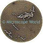
Microscope World has several microscopes set up specifically for wastewater treatment facilities. The most important microorganism that wastewater treatment facilities are looking for is bacteria. In order to view the bacteria, phase contrast is utilized.

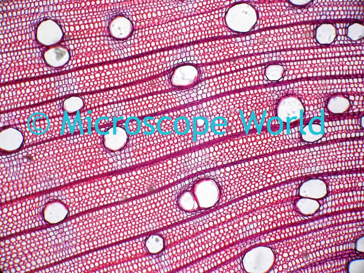
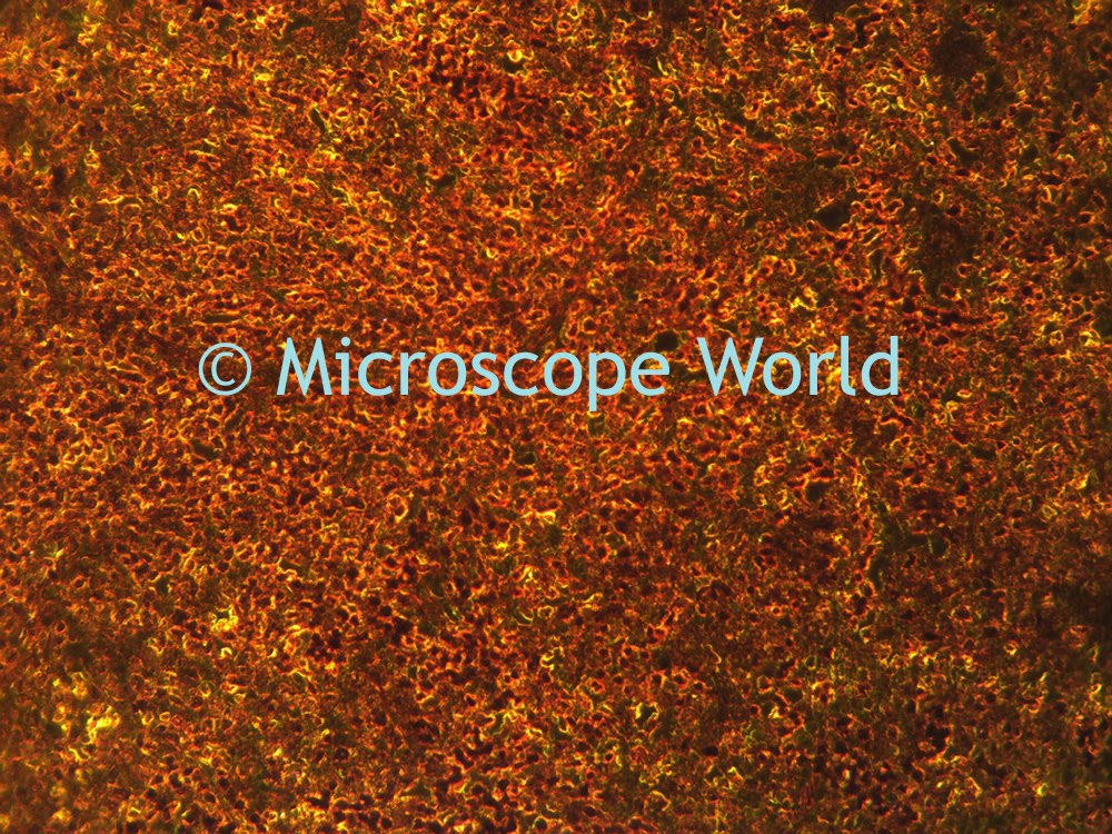
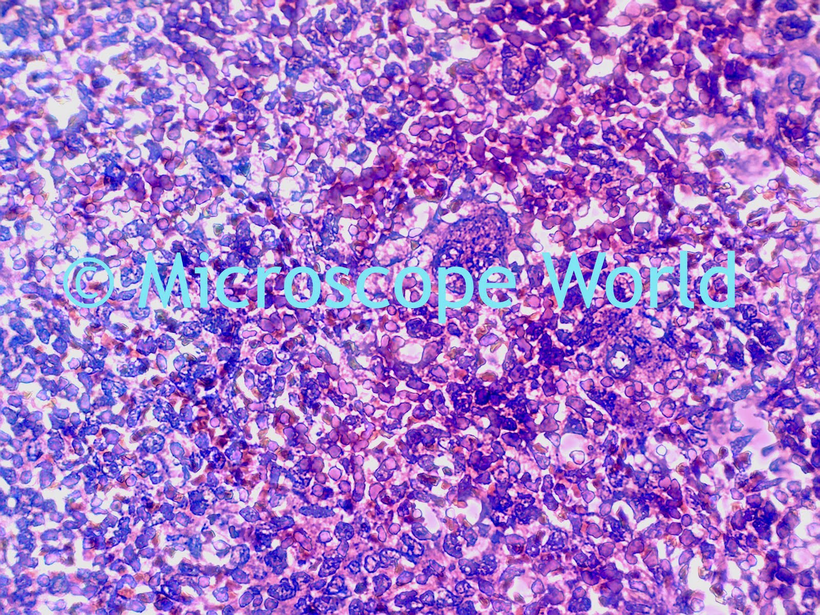
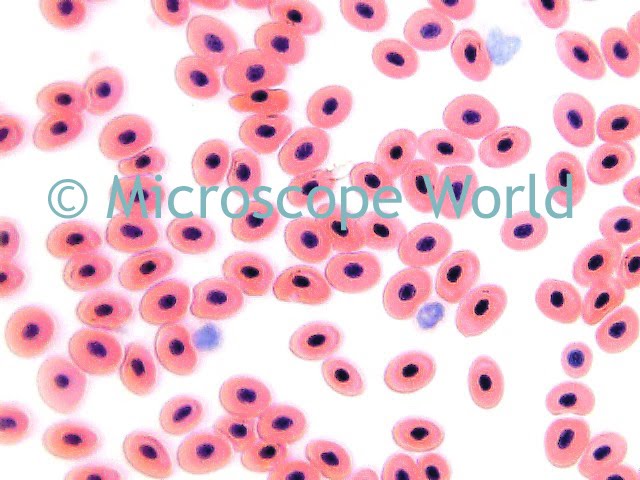
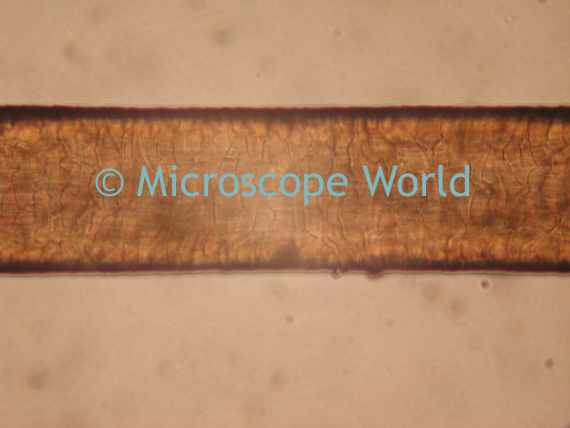
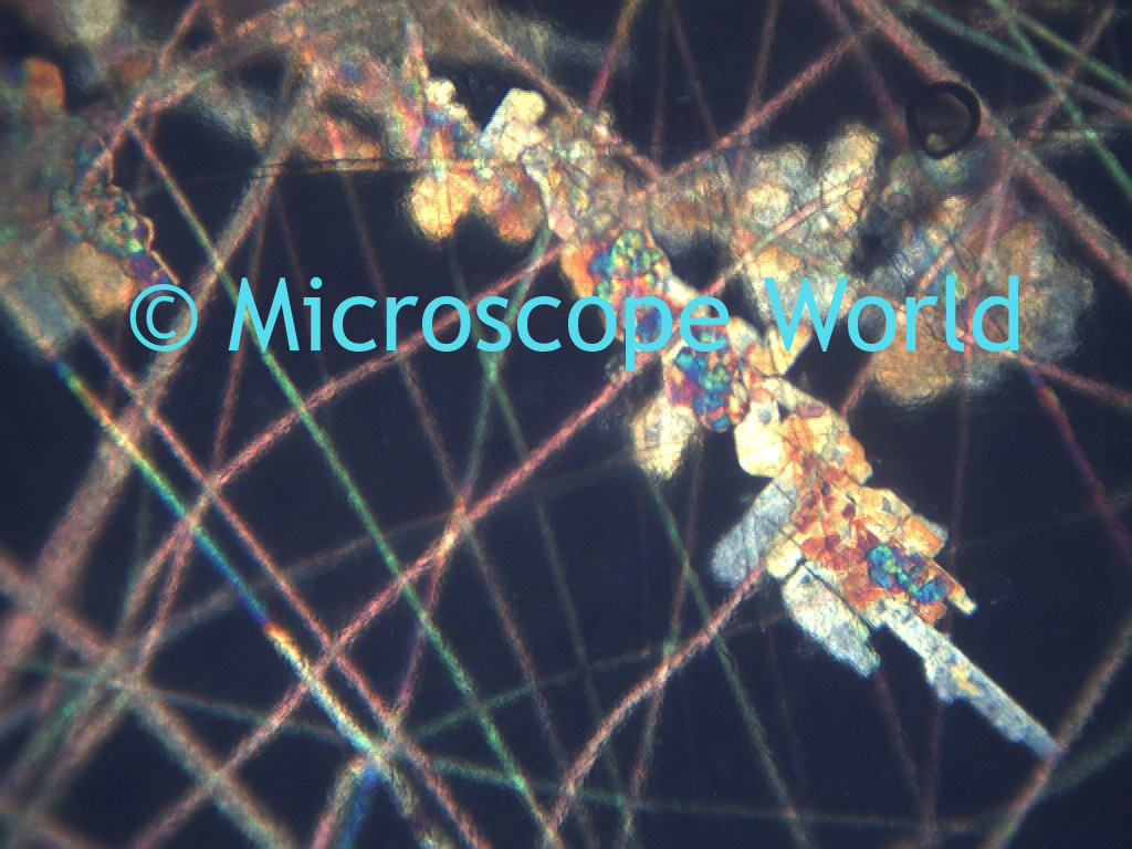
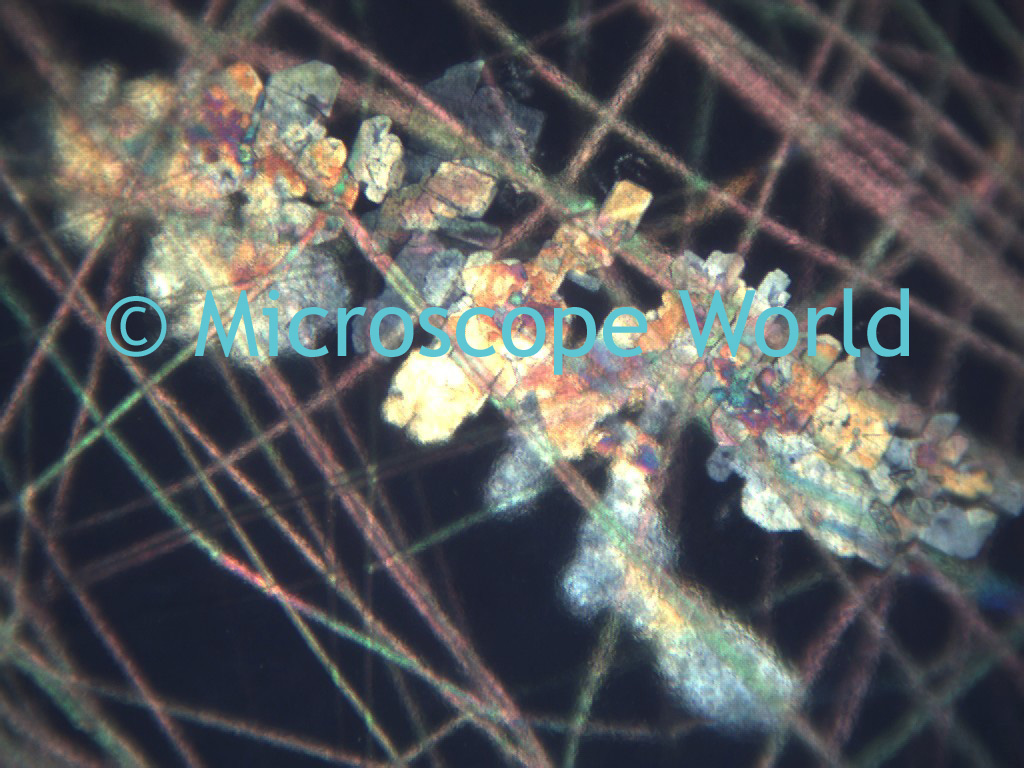
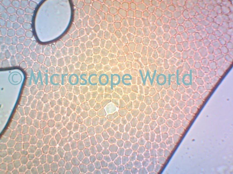
Image of bacteria captured at a wastewater treatment facility using the DC5-163PH digital phase contrast microscope at 400x magnification.

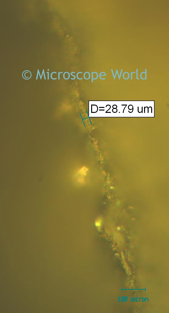
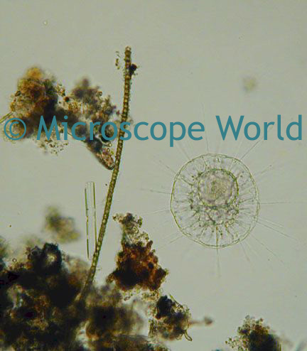
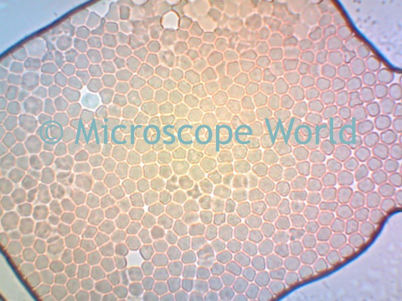
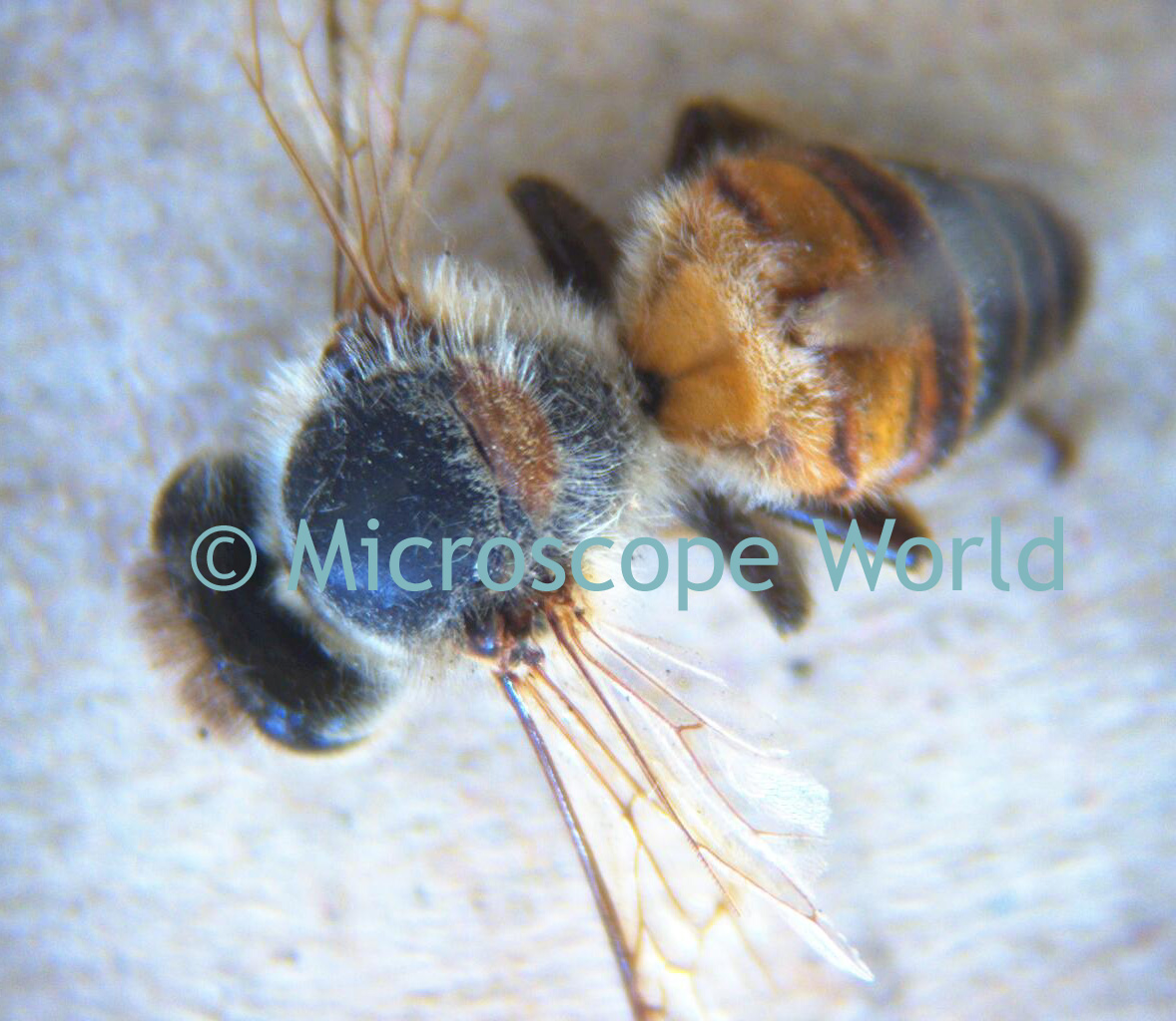
Protozoa are single-celled animals that are the most abundant animals in the world. Wastewater treatment facilities are looking for these creatures. Different forms of protozoa the wastewater treatment plant may be searching for include: amoebas, flagellates, free-swimming ciliates, and carnivore ciliates.

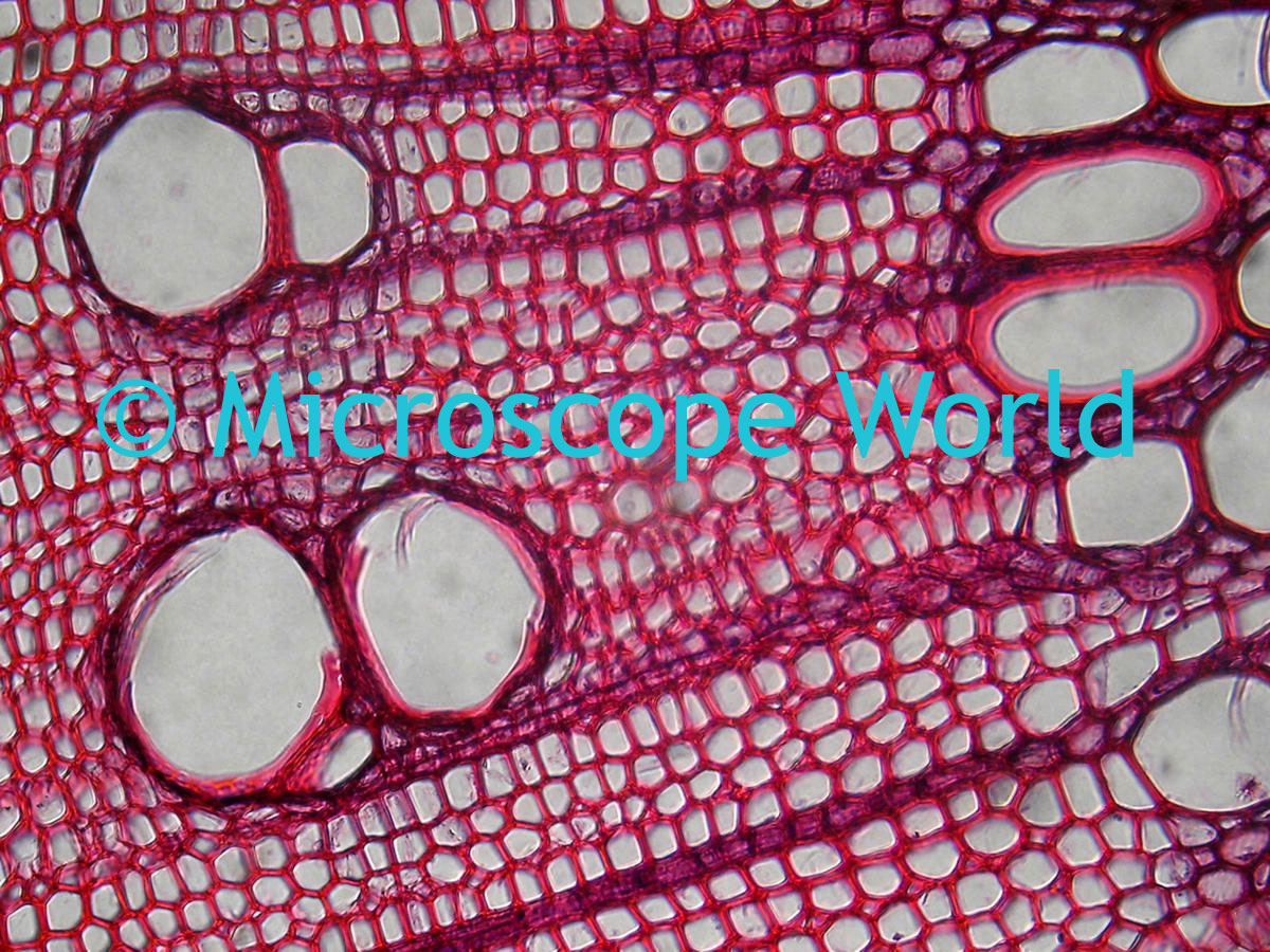
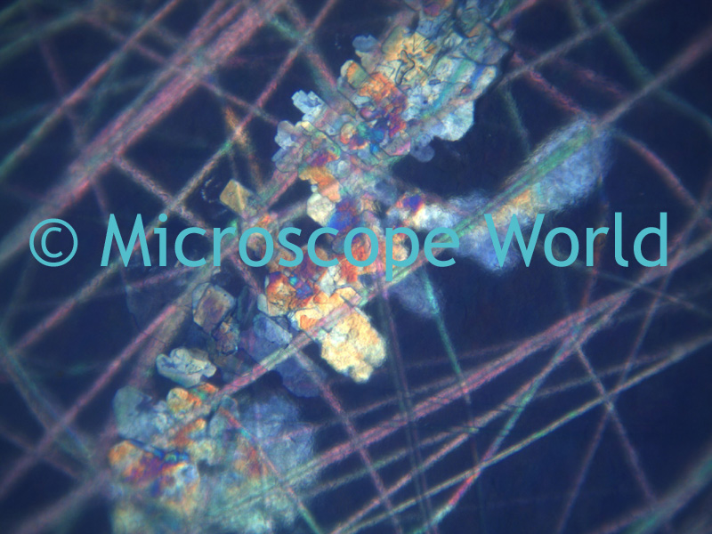
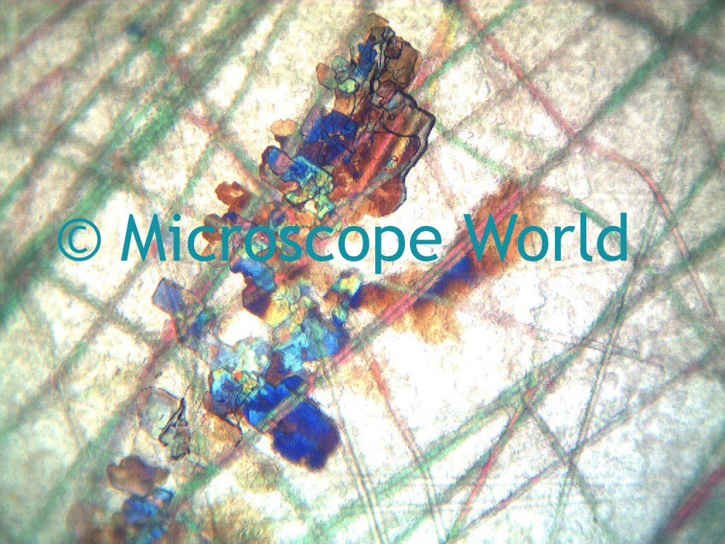
This image of bacteria was captured with the DC5-163PH digital phase contrast microscope at 400x magnification.

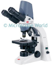
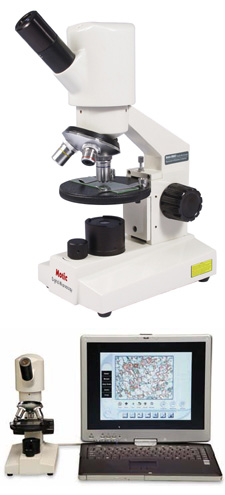
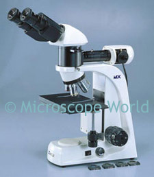
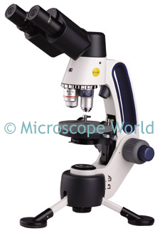
The ability to capture and record images while scanning specimens in wastewater treatment is helpful. The two digital microscopes most commonly used for wastewater treatment are the DC5-163PH and the DMBA310 with phase. If you have any questions regarding phase contrast work please call us at 800-942-0528 and we will be happy to help you select the correct microscope for your needs.

Monday, November 30, 2009

Microscope Project: Mold

Next time you open the fridge and see a moldy piece of bread, cheese or a strawberry, don't throw it out right away. Grab your microscope and learn a little bit first! If you have both a stereo microscope and a compound microscope, you can use each to view the mold.

Starting with the stereo microscope, take a look at the mold. Notice the texture of the mold, it may almost look soft like fabric.

Image of moldy bread at 20x magnification courtesy of King Edward VII School.

Now take the same moldy food and scrape a small piece of mold off with a knife. Place the mold on a blank microscope slide and place a cover slip on top of it. Using a high power microscope, take a look at the mold again. How does it look different? Can you identify any bacteria? Draw a picture of the images that you see and ask your science teacher to help you identify the cells in the mold.

Friday, November 27, 2009

Identifying Particulate Contaminates

Microscope World often helps customers configure a microscope system for identifying particulate contaminates. For example, the standards for European Pharmacopoeia Section 2.9.19, Method II: Microscopic Particle Count Test require the following:
  1. Binocular Microscope
  2. Eyepiece Reticle (Graticule) for specified type
  3. Stage Micrometer with Certification
  4. Mechanical Stage
  5. Episcopic (Reflected) Light Microscope
  6. External Illuminator with Oblique Lighting
  7. 10x Eyepieces with a 10x Objective Lens
The following microscope system was created to meet these standards. The MT7200 reflected light metallurgical microscope, paired with an IMA/USP counting reticle, stage micrometer (NIST certified), and a LED dual pipe illuminator for oblique lighting.

One of our microscope specialists configured this microscope system to meet specific standards.

If you have the need to identify particulate contaminates please give us a call. We would be happy to put a custom quote together to meet your needs.

Tuesday, November 24, 2009

What Can I See With My Microscope?

A compound light microscope has a resolution of about 200nm. A light microscope will allow you to view cells that are alive, fixed or stained.

Bacteria (shown above) is an example of one of the smallest cells that can be seen with a light microscope. Animal and plant cells can be viewed with a light microscope as well.

In order to view small molecules or viruses, a transmission electron or scanning electron microscope is needed. These types of microscopes have a limit of resolution down to 2nm.

Monday, November 23, 2009

Tropism

Tropism is the biological phenomenon that indicates growth or movement of a biological organism (usually a plant) in response to an environmental stimulus.

Photo courtesy of Mark Simmons.

The above tropism is a polarized light microscope image of adipic acid, (CH2)4(COOH)2. The nearly 2.5 billion kg of adipic acid produced each year is mostly used as a monomer for the production of nylon. Other major uses involve polymers. It is a monomer for production of Polyurethane and it is used in making PVC. The image above was captured using a microscope digital camera adapter and a standard digital camera.

Wednesday, November 18, 2009

Microscope Sale

Meiji microscope closeout special. While supplies last, this Meiji microscope system, including fluorescent ring light is $699. High quality Japanese optics. Your choice of 10x, 20x, 30x or 40x magnification. Order online here.

Tuesday, November 17, 2009

Stereo Zoom Microscopes

The Meiji EMZ-13TR stereo microscope was used to capture these images of capacitors.

This image was captured at 75x magnification. In order to achieve this magnification, 20x eyepieces, a 1.5x auxiliary lens and a 1.0x c-mount adapter were used.

This image was captured at 150x magnification.

Monday, November 16, 2009

Hair Science Project

The goal of this science project is to measure hair damage and strength using no hair products and then again after using a variety of different products.

You will need a student microscope with at least 100x magnification. When viewing a single strand of hair, the biological elementary student microscope works well.

Before you begin, find a few friends or family members willing to participate in your science experiment. It is best to use hair that has not been lightened or darkened, as this alters the strength of the hair. Once you have located your subjects, gather a clean strand of hair from each and place it under the microscope. If you can, capture an image of each and label the image with the name, magnification and test # (see example below).

Friend A, 100x magnification, test #1

Friend A, 1000x magnification, test #1

After capturing your images, set the pieces of hair aside in a labeled envelope. Later you will want to pull these out again to test the strength of the hair.

Once you capture the images, have each friend use the same hair care product for a week. Try something that advertises "hair strengthening" or "heals damaged hair". After a week, collect a piece of hair from each person and look at it under the microscope.

Do you notice any difference between the hair from test #1 and test #2? Does the hair care product really make the hair healthier? A damaged piece of hair will have rough edges when viewing it through the microscope. You may want to have the subjects try another week of the hair product and compare a third time.

Finally, take the hair from each session and try to break it by pulling on each end. Do you notice a difference in the strength of the hair that was not treated with any products to those that were?

Did you notice a specific color of hair tended to be stronger than others? If so, you could complete a secondary science project by gathering more samples of similar colors of hair for comparison. If you have great findings from your science project, send us an email, we would love to hear about it!

Friday, November 13, 2009

Nature at 10x and 30x Magnification

Stereo or dissecting microscopes allow you to view nature up close and in detail. Even a simple 20x kids microscope can reveal the stigma of a flower or plant. When viewing items found in nature, a microscope with 10x - 40x magnification is generally best.

This leaf image was captured at 10x magnification,

The same leaf, at 30x magnification.

If you are looking for a good children's microscope that connects to the computer so you can capture and save your nature images, Microscope World recommends the DS2 digital student microscope.

Friday, November 6, 2009

Coin Collecting Microscopes

Coin collecting requires good optics and low magnification in order to fit the entire coin in the field of view. You can view a variety of coin collecting microscopes here.

These images were all captured with the SMZ-168 stereo microscope with 5x eyepieces. The MC1000 microscope camera was mounted over the 5x eyepiece in order to have a lower magnification. This same result could also be obtained using the 10x eyepieces and a 0.5x auxiliary lens.

Tuesday, November 3, 2009

Swift Microscopes

The full line of Swift Microscopes can be found at Swift-MicroscopeWorld. In February of 2010 the micro/macro M3 microscope is being introduced. This microscope allows students to view both micro and macro specimens. The macro setting (20x magnification) is perfect for viewing rocks, plants and insects. The micro magnifications of 40x, 100x and 400x provide standard biological magnifications and allow the student to view protozoans, blood cells, etc.


Pre-orders are being accepted for the Swift M3 microscope, due to ship February 2010.

Thursday, October 29, 2009

Blood Cells

These pictures were captured with the DC5-163 digital microscope. In order to prepare the blood cells, an employee at Microscope World used a clean needle to prick his finger and placed a small drop of blood on a blank glass microscope slide. A cover slip was immediately placed over the small drop of blood and it was given time to dry. While the specimen was drying, the employee used an alcohol swab to clean his pricked finger and put a bandage on it to keep bacteria out it.

These images were captured at 400x magnification. Notice how many cells fill the field of view at 400x magnification! The larger gray areas are air pockets that were caught between the slide and cover slip.

Wednesday, October 28, 2009