Tuesday, September 29, 2009

Microscope Stage Micrometer

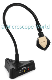
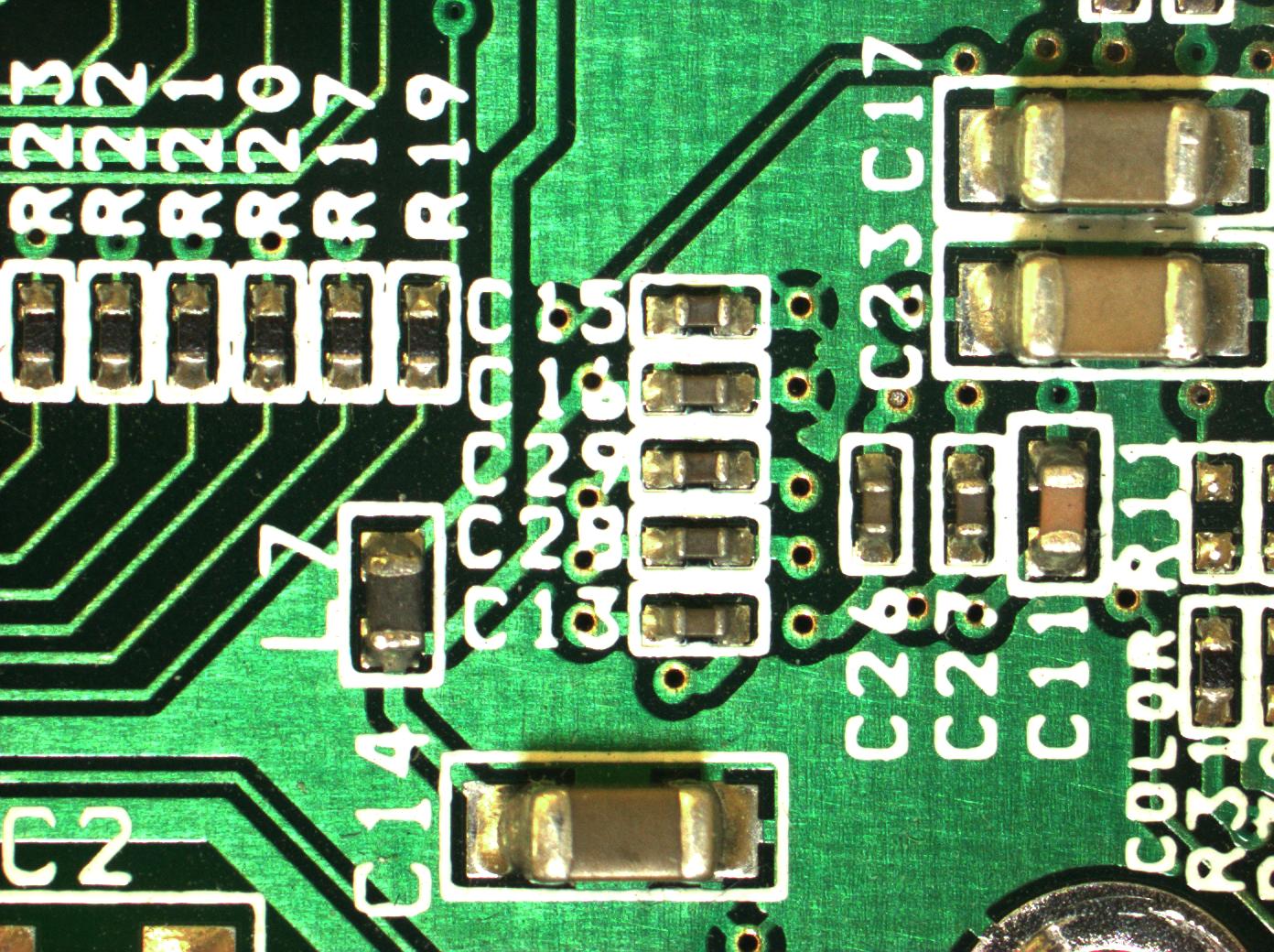
A microscope stage micrometer is used to calibrate an eyepiece reticle. The stage micrometer is a glass slide that has a ruler printed on it. Stage micrometers can be NIST certified for exact points.
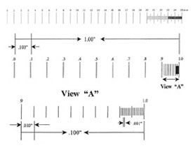
The above is an example of the 1" and 25mm stage micrometer. The top line is what is printed on the stage micrometer. The two lines below explain the divisions on the stage micrometer.

Because each microscope has a slightly different magnification factor, calibrating the eyepiece reticle with a stage micrometer will help you to ensure that your measurements are accurate. Learn more about microscope calibration here.

Monday, September 21, 2009

Darkfield Microscopy

Darkfield microscopy is a simple technique for taking transparent specimens that are not stained and making them clearly visible. Darkfield can be used with either a low power stereo microscope or a high power biological microscope.

Darkfield illumination is a technique where the direct light path is blocked and only oblique light rays are permitted to hit the specimen. This results in an image that appears to almost be back-lit.
An example of a stereo microscope darkfield attachment on the National Optical stereo microscope.

This is the high power darkfield attachment for the National Optical 162 biological microscope. This is a simple plug-in type of darkfield attachment that replaces the condenser.

Darkfield microscopy is often used when viewing blood cells, gems, cell membranes and tissue.

Wednesday, September 16, 2009

Microscope Parts

This image shows the parts of a student microscope. The fine focus helps to get fine-tuned crisp and clear images in your field of view. Some microscopes only have a coarse focus knob, which makes focusing at higher magnifications a bit harder. Notice the rack stop, right behind the objective lenses. This ensures that when looking through the eyepiece while focusing, you do not accidentally run the objective lens into your slide and damage the lens or the slide.

More detail on each of the parts of the microscope listed above can be found here.

Friday, September 11, 2009

Teaching Aids

The video flex camera is an excellent teaching tool. The ability to project a small object onto an LCD projector or a TV monitor so an entire classroom of students can observe makes teachers' jobs easier. The video flex cameras can be used to view maps, insects, they can even be used to project an image from a microscope onto the screen.This is the SwiftCam3 camera. It has the ability to connect to a computer (USB), LCD projector (S-Video) or an older TV (RCA). View live images from the SwiftCam, capture and save images on the computer, or make measurements with the included software!

Wednesday, September 9, 2009

Human Anatomy Microscope Slides

The above image is smooth muscle, captured at 400x magnification with the DC5-163 digital microscope. Smooth muscle is a type of non-striated muscle, found within the walls of hollow organs and in areas such as the bladder, uterus, abdominal cavity, GI tract, respiratory tract and iris of the eye.

Human artery, vein and part of a nerve, captured at 400x magnification with the DC5-163 microscope. Arteries are muscular blood vessels that carry blood away from the heart. A vein is a blood vessel that carries blood toward the heart. A nerve is an enclosed, cable-like bundle of axons (the long, slender projection of a neuron).

The Bacteriology Prepared Slide Kit contains different prepared slides relating to bacteria.

Friday, September 4, 2009

Lumenera Microscope Camera

The Lumenera Infinity2-1 DK1-CCD digital camera has a CCD chip that works well in low-light conditions. High quality images, user-friendly software and affordability make this microscope camera attractive.


Washers captured using a stereo microscope.


Printed circuit board.