Wednesday, November 29, 2017

Fluorescence Microscopy

Fluorescence microscopy is an essential tool in biological and biomedical sciences, as well as in material science. The fluorescence images below were captured using a research microscope with a research microscopy camera.

Green fluorescence microscopy image
Green fluorescence image captured with a research microscope.

Monochrome fluorescence microscopy image
Monochrome fluorescence image captured with a research microscope.

Fluorescnece microscopy image showing multi channel fluorescence.
Multi channel fluorescence microscopy image captured with a research microscopy camera.

For more information on fluorescence research microscopes or research microscopy cameras for your specific application contact Microscope World.

Thursday, November 9, 2017

Tissue Biopsy under the Microscope

A biopsy is a way for a doctor to diagnose a disease. A sample of tissue or cells is removed from the patient by a doctor and sent to a pathologist to be examined under the microscope.

The following images were captured under a pathology microscope during a tissue biopsy.

Microscopy image of tissue biopsy.
Tissue biopsy captured under a pathology microscope.

Microscopy image of tissue captured under a pathologist microscope.
Tissue biopsy captured under a pathology microscope.

Tissue biopsy captured under a pathologist's microscope.
Tissue biopsy captured under a pathology microscope.

Tissue biopsy captured under a pathologist's microscope.
Tissue biopsy captured under a pathology microscope.

To learn more about different types of biopsies click here. For more information on pathology microscopes contact Microscope World.

Thursday, October 19, 2017

Understanding Kidney Disease in Dogs

Kidney failure can cause of death in dogs and there are a number of different ways that kidney disease takes shape in dogs. Veterinarians use a microscope to test for some of the following problems in dogs:
  • Glomerular Disease: This is damage to the kidney filters and can be caused by infections such as Lyme disease or cancer.
  • Pyelonephritis: This is an infection of kidney tissues. If a veterinarian finds this infection, the bacteria that is causing the inflammation of kidney tissues can often be killed.
  • Neophrolithiasis: These are kidney stones. Unlike in humans, kidney stones actually do not typically cause dogs too much pain in the early stages.
  • Ureteral Obstruction with Hydronephrosis: This is a kidney blockage. If a dog has kidney stones and they fragment, they can cause a kidney blockage that will not allow urine to pass and the kidneys will swell and become damaged.
  • Tubulointerstitial Disease: This is damage to kidney tabules, often with an unknown cause. This type of kidney disease can only be determined by using a microscope to examine a kidney biopsy (see image below). 
  • Leptospirosis: This is a bacterial infection and can cause kidney disease along with organ problems in both people and dogs. Infection can be caused by contact with urine or other bodily fluids from the infected dog.
  • Toxins: These are medications, household chemicals, and even ordinary food like grapes or raisins. If your pet ingests toxins contact ASPCA Poison Control immediately.
  • Cancer: The good news is that kidney cancer is not common in dogs. The bad news is that treatment options for kidney cancer in dogs is quite limited. If only one kidney is affected it can be removed with a positive outcome. If the cancer is benign or has not spread the outcome can be good as well.
  • Amyloidosis: This is a protein issue with the kidneys where protein deposits replace normal tissue. 
  • Hereditary: Some purebred dogs have a higher rate of kidney disease than others. Additionally, some young dogs will fail to develop normal kidneys.
The image below is of a dog kidney and was captured using a veterinary lab microscope with a microscope camera.

Dog kidney captured under a veterinarian's microscope.
Dog Kidney under the Veterinary Microscope

For more information about veterinarian microscopes contact Microscope World.

Friday, September 22, 2017

Lily of the Valley under the Microscope

Convallaria is a genus of flowering plants and includes Lily of the Valley. This is a plant that is sweetly scented and highly poisonous and is native throughout the cool temperate Northern Hemisphere in Asia and Europe.

Convallaria majalis is an herbaceous perennial plant that forms extensive colonies by spreading underground stems and rhizomes.

The images below of Convallaria were captured using the Zeiss Primostar microscope.

Microscopy image of lily of the valley captured under a Zeiss Primostar microscope.
Lily of the Valley rhizome with concentric vascular bundles, captured under Zeiss Primostar microscope at 400x.

Zeiss Primostar microscope image of a lily under the microscope at 100x magnification.
Lily of the Valley rhizome with concentric vascular bundles, captured under Zeiss Primostar microscope at 100x.

Contact Microscope World with questions regarding Zeiss microscopes or capturing images with your microscope.

Tuesday, September 12, 2017

Chromosomes under the Microscope

A chromosome is a DNA molecule with part or all of the genetic material of an organism. Chromosomes are only visible under a light microscope when the cell is undergoing the metaphase of cell division. During metaphase, when a chromosome is in its most condensed state, the X-shape structure is called a metaphase chromosome.

The images below were captured under the Zeiss Primostar microscope.

Zeiss Primostar microscope images of chromosomes captured at 100x.
Chromosomes captured under the Zeiss Primostar microscope at 100x.

Zeiss Primostar microscope image of chromosomes captured at 400x.
Chromosomes captured under the Zeiss Primostar microscope at 400x.

Contact Microscope World with questions regarding microscopes and different applications.

Tuesday, August 22, 2017

Plant Cell Images from Student Microscope

The plant cell microscopy images below were captured with the Richter Optica UX-1 student microscope using Plan Achromat objective lenses and the high definition HD camera with monitor.

Microscopy image of Monocot captured under a student microscope by Microscope World.
Monocot captured under the student microscope.

Plant cells captured using the microscope HD camera with monitor.

Contact Microscope World with questions regarding microscopes or microscopy cameras.

Wednesday, August 16, 2017

Muscle under the Microscope

The human body has 3 types of muscle in it:
  • Skeletal muscle
  • Smooth muscle
  • Cardiac muscle
Skeletal muscle covers the skeleton and gives the body shape. Skeletal muscle is the type of muscle that is most often thought of when the term "muscle" is used.

Muscle under the microscope captured with a PAXcam microscope camera.
Skeletal muscle under the microscope captured with a PAX-cam microscope camera.

Microscopy image of smooth muscle.Smooth muscle (shown at left) is found in walls of hollow organs such as the stomach. Smooth muscle tissue tends to demonstrate greater elasticity than other muscles. It is also found in the urinary tract and digestive tract.


Cardiac muscle is only found in the heart and it is different from the other muscles in the body because it does not get tired. Learn more about the human cardiac muscle here.

Microscopy image of human heart captured at 400x magnification.
Human cardiac muscle captured under the RB30 microscope with a 5mp microscope camera.


Each of the 3 types of muscles in the human body plays an important function in every day life. To learn more about healthy muscles and the roles they play in the human body visit NIH here.

Tuesday, July 18, 2017

Marmalade Hoverfly under the Microscope Lens

Harald K. Andersen of Steinberg, Norway captured the image below of a marmalade hoverfly. His setup included using the Mitutoyo M Plan Apo 5x Objective lens, connected to his Canon EOS 7D camera.

The marmalade hoverfly (Episyrphus balteatus) is a relatively small hoverfly of the Syrphidae family, widespread through the Palaearctic region which covers Europe, North Asia and North Africa. The upper side of the abdomen is patterned with orange and black bands. These insects often form dense migratory swarms, which may cause panic among people for their resemblance to wasps.

Harald captured this image using a 100 image stack (Canon EF 70-200 f/2.8).

Mitutoyo lens on Canon EOS 7D Camera
Mitutoyo M Plan Apo 5x Objective on Canon EOS 7D

Microscopy image setup for photography
Harald K. Andersen's studio

Harald K. Andersen image of marmalade hoverfly in 100 image stack.
Marmalade hoverfly courtesy of Harald K. Andersen (100 image stack)

Contact Microscope World with questions regarding objective lenses.

Wednesday, June 28, 2017

Microscope Ergonomic Work Station

A proper ergonomic work station when using your microscope can reduce the risk of occupational injuries. UCLA Ergonomics suggests the following tips to maintain a safer environment when using microscopes:
  • Sit in the proper position keeping your back straight and your shoulders back. Distribute your body weight evenly on both hips, and keep your feet flat on the floor.
  • Arrange your work space so that it is close to you.
  • Ensure there is proper padding if leaning on hard surfaces.
  • Work with elbows in close proximity to the body.
  • Work with wrists in a straight and neutral position.
  • Adjust and/or elevate your chair, workbench, or microscope as needed to maintain an upright head position.
  • Adjust microscope eyepieces or mount the microscope on a 30° stand for easier viewing.
  • Repair and clean microscopes regularly. Learn how to clean lenses here.
  • If possible, spread microscope work throughout the day and between several people. 
  • Schedule works breaks. Every 15 minutes, close your eyes or focus on something in the distance. Every 30 minutes get up to stretch and move.
Microscopy is a demanding and rewarding field to work in. By maintaining an ergonomic work space you can ensure years of healthy and pain-free work.

Sources:
"Tips for Microscopy." Laboratory Ergonomics. Ergonomics.ucla.edu, 2012. Web. 27 June 2017.
"Posture for a Healthy Back." Articles. Health. Clevelandclinic.org, 2017. Web. 27 June 2017.

Wednesday, June 21, 2017

Persimmon Endosperm

Endosperm is a tissue produced inside seeds following fertilization in most flowering plants. It surrounds the embryo and provides nutrition in the form of starch, though it can also contain oils and protein. This can make endosperm a good source of nutrition in the human diet. The image below is of a persimmon endosperm captured under the microscope using the Lumenera Infinty 2-1 color microscopy camera.

Persimmon endosperm captured under the microscope with Infinity 2-1 microscopy camera.
Persimmon Endosperm under the Microscope captured with Infinity 2-1 Camera

Contact Microscope World with questions regarding microscopes or microscopy cameras.

Wednesday, June 14, 2017

Microscope Magnification versus Resolution

In microscopy there are two concepts that many people often think of as a single concept, but they are very different. These two concepts are magnification and resolution. From a technical perspective, resolution is a quantified concept that is defined by the numerical aperture (NA) rating of the objective lenses of the microscope. Numerical Aperture is a number that expresses the ability of a lens to resolve fine detail in an object being observed. Magnification is simply how much an image is enlarged.

Below are two images of the same small printed part with text on it. The first image was captured using a stereo microscope with a lens that has NA of 0.10. This image was captured at 90x. Notice in the image below captured with the stereo microscope it is very hard to even read any of the printed text on the circuit. It should also be noted that it took nearly two hours to capture an image of this quality.

Image of a circuit captured under a stereo microscope at 90x magnification.
Stereo Microscope image captured at 90x, NA 0.10

The next image was captured using a metallurgical microscope with a lens that has NA of 0.30 and a magnification of 100x. This image took a few minutes to capture. The magnifications of the two captured images are similar however, notice how much easier it is to read the printed letters in the image that was captured with the metallurgical microscope. That ability is due to better resolution, which was obtained because of a higher numerical aperture of the lens used.

Metallurgical microscope image of a circuit captured at 100x magnification showing high quality resolution.
Metallurgical Microscope image captured at 100x, NA 0.30

Contact Microscope World with questions regarding NA, resolution, magnification or any other microscopy related questions.

Thursday, June 8, 2017

Corn Grain Under the Microscope

Corn is a large grain plant that was first domesticated by people in southern Mexico nearly 10,000 years ago. The ears of corn are actually fruit that produce the kernels (seeds) that people eat. Total production of corn worldwide surpasses that of wheat or rice. However, this is not all used for food. Corn is also used for producing ethanol, animal feed, corn starch and corn syrup.

The United states produced 361 million tons of corn in 2014, followed by China with 215 million tons and Brazil with 80 tons.

The images below of a cross section of kernel of corn were captured under the microscope using the Lumenera Infinity 3-3UR microscope camera.

Microscopy image of kernel of corn.
Grain of corn captured under the microscope with Infinity 3-3UR microscopy camera.

Microscope image of a kernel of corn.
Grain of corn captured under the microscope with Infinity 3-3UR microscopy camera.

Contact Microscope World for more information on Lumenera cameras or microscopes.

Friday, June 2, 2017

Middle School Microscopes

Microscope World offers several middle school microscopes that are perfect for kids exploring science!

MDS1 Middle School Microscope

Richter Optica MDS2 Middle School Microscope is a cordless and rechargeable microscope.
The Richter Optica MDS1 middle school microscope has several features that make it appealing to middle school classrooms and parents looking for a first microscope for their kids.
  • Cool, LED illumination (the bulb lasts a LONG time)
  • Carrying handle - perfect for younger students
  • Coarse & fine focusing, a must when viewing 400x
  • Cordless & Rechargeable - run the microscope corded or cordless
  • Retractable 40x lens helps teachers protect slides and the lens.

MDS2 Middle School Microscope

Richter Optica MDS2 Middle School Microscope has the ability to add a mechanical stage.
The Richter Optica MDS2 middle school microscope is a step up from the MDS1 in that it allows for the attachment of a mechanical stage. Here are a few features of the MDS2 middle school microscope. (Image shown includes optional mechanical stage).
  • Cool, LED illumination (the bulb lasts a LONG time) 
  • Coarse & fine focusing, a must when viewing 400x
  • Corded microscope
  •  Retractable 40x lens helps teachers protect slides and the lens.
  • Ability to add a mechanical stage, providing more precise control over X-Y movements of slides. 


MW2-HB4 Middle School Microscope

MW2-HB4 cordless middle school microscope from Microscope World.
The MW2-HB4 middle school microscope has a bit of a wider frame, providing added stability to the microscope.  A few features of this middle school microscope include the following:
  • Cool, LED illumination (the bulb lasts a LONG time) 
  • Coarse & fine focusing, a must when viewing 400x
  • Cordless & Rechargeable - run the microscope corded or cordless
  • Retractable 40x lens helps teachers protect slides and the lens.

Contact Microscope World with questions regarding middle school or student microscopes. 

Wednesday, May 31, 2017

Motic Images Software Calibration Tutorial

Motic Images software is included with all Motic microscopy cameras. The software allows for image capture, annotation and one of the most commonly used features of the software: making measurements! However, before you can accurately make measurements with the Motic Images software, it is important to calibrate your microscope with the software first.

This video walks you through the steps of Motic Images microscopy software calibration.


Contact Microscope World with questions regarding Motic Images software or Motic microscope cameras.

Wednesday, May 17, 2017

Motic Panthera Microscopes

Microscope World is proud to carry the new line of Motic Panthera microscopes. Panthera is Motic's new upright microscope line. There are several Panthera microscopes available.

Motic Panthera S school microscope with fixed koehler illumination can run off a battery pack if needed.Panthera S School Microscopes

The Panthera S is a school model. This microscope has Plan SC Achromat objective lenses and an extremely efficient low power illumination that allows the microscope to run on a mobile battery pack for several hours. The Panthera S is a fixed Koehler LED microscope for the educational market and is available in both binocular and trinocular.


Motic Panthera U Microscope with LightTracer illumination feature.

Panthera U University Microscopes

The Panthera U microscopes were created for University use. The Motic Panthera U microscopes have Plan UC Achromat higher quality objective lenses than the Panthera S. The microscope has fixed Koehler 3W LED illumination, and a larger stage than the Panthera S model. The microscope features Motic LightTracer, a digital illumination control. The microscope has a digital intensity knob with coded LED nosepiece that controls the illuminator to offer information on the current light intensity of each objective. Once the microscope is calibrated for each objective lens, the user does not need to adjust the illumination intensity when changing the microscope magnification. The Panthera U is available in binocular or trinocular.

Motic Panthera C Classic Microscopes

Panthera C Classic Microscopes

The Moitc Panthera C microscopes were created for the traditional microscope user. This is a classic all-around microscope that provides both Halogen and LED full Koehler illumination with manual light management. The Panthera C has Plan UC Achromat objective lenses and this microscope includes an integrated USB camera power port (it does not include a camera) and an LED light intensity illumination indicator in the nosepiece. The Panthera C classic microscopes are available in binocular or trinocular.
Motic Panthera L Life Sciences Digital Microscope

Panthera L Life Sciences Digital Microscopes

The Panthera L Life Sciences digital microscopes have built-in digital capabilities. This microscope has Plan UC Achromat objective lenses and Halogen and LED full Koehler illumination. Motic LightTracer provides a digital intensity knob with coded LED nosepiece that controls the illuminator to offer information on the current light intensity of each objective. Once the microscope is calibrated for each objective lens, the user does not need to adjust the illumination intensity when changing the microscope magnification. Digital connection by HDMI, USB, WiFi or RJ-45 allows direct image projection to a monitor or the use of the Panthera App on a mobile device. The Panthera L microscope is available in only one binocular model with a built-in camera.

Motic Panthera HD Digital Microscope

Panthera HD Digital Microscope

The Motic Panthera HD Digital microscope has all the same features as the Panthera L, but it is meant to only be used as a digital microscope. The Panthera HD does not have any eyetubes or eyepieces.


Contact Microscope World with any questions regarding the Motic Panthera microscopes.
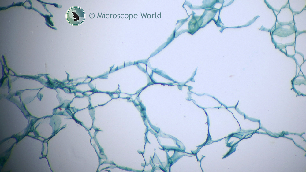
Friday, May 5, 2017

Tips for Cleaning Microscope Optics

There are several things to watch out for when cleaning microscope optics. Follow some of these tips when cleaning your microscope lenses to ensure you do not scratch or damage them.

    Tips for cleaning microscope lenses.
  • When cleaning a microscope eyepiece, start out by using a dust blower to remove dust particles.
  • Never wipe lenses with dry tissue as it can scratch the lenses.
  • Do not use any solvents before trying distilled water. A thin film of distilled water can always be created by breathing on the surface of the lens.
  • Do not use abrasive materials such as a dry linen cloth to clean microscope lenses as they may scratch the surface.
  • Older microscopes should not be cleaned using ethanol, diethyl ether or acetone.
  • Use Clean Tip swabs rather than Q-Tips for cleaning.
  • Refrain from using optical spray cans containing pressurized liquid air to clean lenses. These can leave a residue that is difficult to remove.
  • Do not use acids or ammonia to clean microscope objective lenses.
  • Do not try to clean internal optical surfaces, cameras or adapter optics. Contact a microscope service technician if you need to clean these items.

Contact Microscope World regarding microscope service and repair.

Tuesday, May 2, 2017

Pap Smear Under the Microscope

A Pap smear is a screening procedure used to test for cervical cancer. The test looks for precancerous or cancerous cells on the cervix. The Pap smear was named after Georgios Papanikolaou, the doctor who determined that the test was a helpful way to detect cervical cancer.

This is a slide from a Pap smear that was captured under the RB20-D digital lab microscope at 400x magnification.

Microscopy image of a pap smear captured at 400x under the U2D microscope.
Pap smear captured under the RB20-D Digital Lab Microscope at 400x.

Contact Microscope World with microscopy questions.

Thursday, April 27, 2017

Sickle Cell Anemia under the Microscope

Sickle cell anemia is an inherited form of anemia. It is a condition in which there are not enough healthy red blood cells to carry adequate oxygen throughout the body.

Normally, red blood cells are flexible and round, moving easily through blood vessels. When a person has sickle cell anemia, the red blood cells become rigid and sticky and are shaped like sickles or crescent moons. These irregularly shaped cells can get stuck in small blood vessels, which can slow or block blood flow and oxygen to parts of the body.

Microscopy image of sickle cell anemia at 40x magnification.
Sickle Cell Anemia under the microscope at 40x.

Symptoms of sickle cell anemia include fatigue, episodes of pain (when blood cells are being blocked), swelling of hands and feet, delayed growth, and vision problems.

Microscope World image of sickle cell anemia under the microscope at 100x.
Sickle Cell Anemia under the microscope at 100x.

There is no cure for most people with sickle cell anemia. Bone marrow transplant or a stem cell transplant is the only potential treatment, but it is only used in children under age 16 because of the increased risk for older patients. Treatment for sickle cell anemia is usually aimed at avoiding crises, relieving symptoms and preventing complications.

These images are of sickle cell anemia cells that were captured under a lab microscope using a 5 megapixel microscopy camera.

Microscope World image of sickle cell anemia captured at 400x.
Sickle Cell Anemia under the microscope at 400x.

Learn more about sickle cell anemia here.

Contact Microscope World with questions regarding microscopes and microscopy cameras.

Tuesday, April 25, 2017

High School Binocular Microscope

The Richter Optica HS-3B is a versatile high school compound microscope. A few features make this microscope one of the most popular microscopes across high schools in the United States.
  • Binocular eyetubes with WF10x eyepieces.
  • 40x, 100x, 400x, 100x magnification.
  • Retractable 40x and 100x (oil) objective lenses avoid damage if moved into contact with the stage.
  • Frame provides easy carrying handle, with cord storage behind microscope.
  • Operates corded or cordless.
  • Rechargeable batteries.
  • LED light is cool and provides long-lasting bulb.
  • Mechanical stage is attached and locked on.
  • Coarse and fine focusing allows for a crisp and clear image.
  • 100~240V allows for use in all countries.
  • Microscope is also available in pared down 400x magnification model, HS-3B-3


HS-3B Student Microscope
Richter Optica HS-3B Microscope

You can view all the Richter Optica microscopes and accessories here. Contact Microscope World for a quote.

Tuesday, April 18, 2017

Mouse Intestine under the Microscope

The images below of a mouse intestine were captured using a Lumenera Infinity 3-1 microscope camera. This is a cooled, CCD camera that is great for low-light situations or where a high dynamic range is required. This 1.4 megapixel camera is ideal for the following applications:
  • Fluorescent Microscopy
  • Green Fluorescent Protein Applications
  • Fluorescent In Situ Hybridization
  • DNA Analysis
  • Live Cell Imaging
  • Brightfield, Darkfield, DIC/Phase Contrast
  • Near IR Applications
  • Histology, Pathology and Cytology
  • Forensic Analysis
  • Semiconductor Inspection
  • Metallurgical Microscopy 
Mouse intestine under the microscope captured with Lumenera Infinity 3-1 CCD camera.
Mouse intestine captured with the Monochrome Infinity 3-1 microscopy camera.

Mouse intestine captured with the Color Infinity 3-1 microscopy camera.

Infinity 3-1 microscopy camera image of mouse intestine under the microscope using fluorescence.
Mouse intestine captured with the Color Infinity 3-1 microscopy camera.

Contact Microscope World for more information on microscopy cameras.

Friday, April 14, 2017

Hair Under the Microscope

Harald K. Andersen of Steinberg, Norway captured these images of hair under the microscope. The first image is of a single strand of hair captured through the microscope at 20x magnification. The hair was dyed for the project.

Hair captured under the microscope.
(C) Harald K. Andersen. Single strand of hair under the microscope.

In order to capture the next image Andersen stacked together 480 images using a software called Zerene Stacker.

Hair captured under the microscope.
(C) Harald K. Andersen. 480 stacked images of a single strand of hair under the microscope.

Andersen mentioned that the hardest part of the project was tying the knot in the hair! A big thanks for sharing the images with Microscope World!

Friday, April 7, 2017

Tonsils under the Microscope

The palatine tonsils are two lymphatic masses, located at the back of the throat on each side of the oral pharynx. The tonsils play a role in protecting the body against respiratory and gastrointestinal infections.

Each tonsil consists of a network of crypts (pits) that store cells used to fight infection. The tonsils contain B cells, a type of white blood cell that fights infections. They also produce antibodies against polio, streptococcal pneumonia, influenza, and numerous other infections. Antibodies are proteins that help the body identify and attack harmful invaders.

The tonsils also contain several types of T cells, which are white blood cells that destroy cells infected with viruses and help the body build immunity to infectious organisms.

The images below are cross sections of Palatine Tonsils that were captured using a biological lab microscope and a microscopy camera.

Microscopy image of tonsil under the microscope at 40x captured by Microscope World.
Palatine tonsil under the compound microscope at 40x.

Microscope World image of palatine tonsil cross section under the microscope at 100x.
Palatine tonsil under the compound microscope at 100x.

Microscope World image of palatine tonsil cs captured at 400x.
Palatine tonsil under the compound microscope at 400x.

Contact Microscope World with any questions about microscopy or capturing digital images under the microscope.

Tuesday, April 4, 2017

Worms found in Cows under the Microscope

Bunostomum phlebotomum is a species of worms (nematodes) that are found in the small intestine of calves. The worms are stout and measure 1-3cm in length. Young animals are most commonly affected and only several hundred worms can cause death. Adults often carry worms but do not show any symptoms.

Diarrhea is the primary symptom in the cows along with signs of anorexia, lethargy and weight loss. Hypoproteinaemia, anaemia and dehydration often occur. Skin involvement due to larvae penetration appears as lesions especially on the feet and limbs. Analysis of fecal samples under a microscope enables eggs to be found, or adult worms can be visualized in the small intestine when an autopsy is performed.

Veterinarians and ranchers can use Anthelmintics (antiparasitic drugs) for effective treatment of the infection. Preventative antiparasitic treatment may help prevent the disease as well as pasture management to avoid egg and larvae accumulation.

The image below is Bunostomum phlebotomum that was taken from a calf in Texas. Images were captured using a 3 megapixel microscopy camera on the RB20 Lab Microscope.

Microscope World image of bunostomum phlebotomum under the microscope at 100x.
Bunostomum Phlebotomum (worm) taken from a cow's intestine, examined under the RB20 Microscope at 100x.

Microscope World image of Bunostomum phlebotomum under the microscope at 400x.
Bunostomum Phlebotomum (worm) taken from a cow's intestine, examined under the RB20 Microscope at 400x.