Thursday, August 25, 2022

ZEISS Stemi 305 Stereo Microscopes

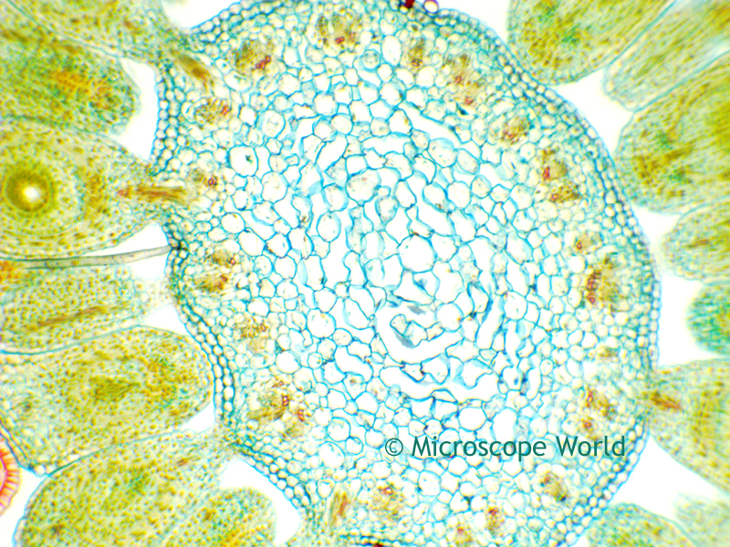
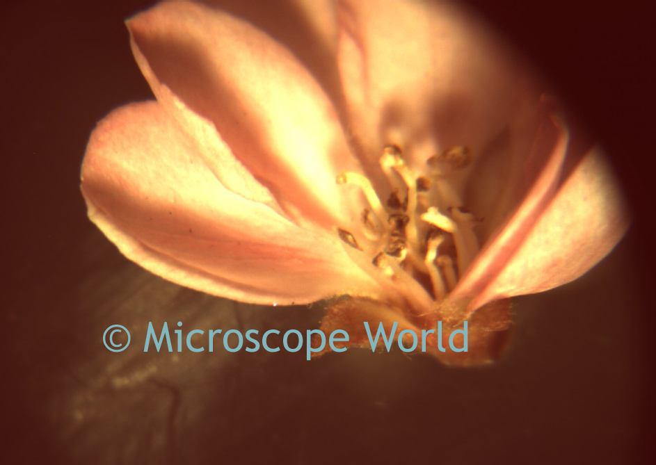
The ZEISS Stemi 305 microscopes offer a variety of illumination options on a clean, compact stand. It's easy to add a camera for digital microscopy. View images of samples captured with the ZEISS Stemi 305 microscopes here.

Darkfield Stereo Microscopy

ZEISS Stemi 305 Stereo Microscope Snails using Darkfield
Snails under Stemi 305 K EDU Darkfield

The ZEISS Stemi 305 K EDU stereo microscope provides brightfield, darkfield, and oblique illumination. 

8x - 40x Zoom Magnification

Adjustable spot illuminator.

Near Vertical Illumination.

Easily switch between brightfield and darkfield.


ZEISS Stemi 305 K EDU Microscope

Material Stereo Microscope

The ZEISS Stemi 305 MAT provides ESD protection, 48 LED Ring Light, and 8x - 40x zoom magnification. Near vertical illumination is controlled with the stand.


ZEISS Stemi 305 MAT Microscope

Lab Stereo Microscope

The ZEISS Stemi 305 LAB microscope is ideal for working in a laboratory and observing or preparing bio specimens. The mirror-based transmitted light is perfect for viewing transparent specimens. Near vertical illumination, dual spot illuminators, and brightfield / darkfield illumination all provide options for viewing samples in an ideal setting. 8x - 40x Zoom Magnification.


ZEISS Stemi 305 LAB Microscope

You can view all of the ZEISS Stemi 305 microscopes here. Have questions about what stereo microscope is best for your needs? Contact Microscope World.

Tuesday, October 16, 2018

Fluorescence Microscopy

This beautiful video of fluorescence microscopy was captured using the HDCAM5 microscope camera.


Contact Microscope World with questions regarding fluorescence microscopy or capturing images and video of fluorescence microscopy.

Wednesday, October 10, 2018

Protozoan Parasite under the Microscope

OE (Ophryocystis Elektroscirrha) is a protozoan parasite that infects butterflies in the Danaus species group, those that host on milkweed. This photo of some Monarch butterfly abdominal scales was taken by Rob Wood with a Canon 80D DSLR through a Richter Optica HS-3 Biological microscope, at 400X. The large objects are scales, while the football-shaped objects are OE spores. Each spore contains 8 protozoa.

Ophryocystis Elektroscirrha captured under the Richter Optica HS-2 microscope.
Ophryocystis Elektroscirrha under the HS-3 microscope.

Thank you Rob for sharing this photo with Microscope World!

Monday, September 17, 2018

Woody Stem Under the Microscope

Below is a woody stem cross section captured under the Richter Optica UX1 cordless student microscope captured at 400x magnification. This image was captured using the DCM5 microscope camera (5.2 megapixels).

The woody stem produces wood as its structural tissue - typically either a tree, shrub or liana.

Woody stem cross section under the Richter Optica UX1 microscope at 400x.
Woody stem c.s. under the UX1 cordless microscope with DCM5 camera.

Tuesday, May 22, 2018

pco Cameras

Microscope World is proud to offer pco scientific cameras. The pco cameras are known for their low readout noise, high frame rates and exposure times that range from nanoseconds to hours. UV and NIR detection and high dynamic range are just some of the pco cameras' unique core competences.

Microscope World offers the pco.panda 4.2 camera.
pco.panda 4.2 Camera


The pco.panda 4.2 camera combines revolutionary sCMOS technology in a compact design. High quantum efficiency (up to 80%) and low readout noise make this camera suitable for countless applications. The USB 3.1 interface enables ultra-speed data transfer and direct power via the USB cable, making external power supplies redundant. Frame rate = 40 fps @ full resolution 2048 x 2048, 80 fps @ 2048 x1024.

pco.edge camera from Microscope World
The pco.edge 3.1 camera has a sCMOS sensor and is designed for users who require high resolution and high frame rates. This USB3 camera is available in color or monochrome. 50 fps @ full resolution 2048 x 1536, 75 fps @ 1280 x 1024, 160 fps @ 640 x 480.

The pco.edge 4.2 camera has sCMOS technology and can be optionally upgraded with a water cooling system. This camera has high quantum efficiency at up to 82% at peak. 100 fps @ RS fast scan 2048 x 2048, 189 fps @ fast scan 1920 x 1080, 420 fps @ fast scan 640 x 480 when using camera link.

The pco.edge 4.2 LT camera has sCMOS technology and USB3 output. High quantum efficiency of up to 82% at peak. 40 fps @ full resolution 2048 x 2048, 80 fps @ 1280 x 1024, and 170 fps @ 640 x 480.

The pco.edge 5.5 camera has sCMOS technology and can be optionally upgraded with a water cooling system. Quantum efficiency of  > 60% @ peak. With camera link, the frame rates are high: 100 fps @ RS/GR fast scan 2560 x 2160, 201 fps @ fast scan 1920 x 1080, 450 fps @ fast scan 640 x 480.

Wednesday, May 16, 2018

Fluorescence Microscopy

Fluorescence microscopy is a tool used in biology, material science, and biomedical sciences. The fluorescence microscope can reveal the presence of a single molecule. The basic function of a fluorescence microscope is to irradiate the specimen with a desired and specific band of wavelengths, and then to separate the much weaker emitted fluorescence from the excitation light.

The fluorescence microscopy images shown below were captured using the Fein Optic RB30-GFP fluorescence microscope and the Lumenera Infinity 3S-1UR microscope camera.

Fein Optic RB30-GFP fluorescence microscope image.
Fluorescence microscopy image captured using a 60x objective lens.

Fein Optic RB30-GFP fluorescence microscope image from Microscope World.
Fluorescence microscope image, 600x.

Contact Microscope World with questions regarding fluorescence microscopy.

Thursday, April 19, 2018

Spring Flowers under the Microscope

In the spirit of welcoming spring, below are some photos of flowers captured under the biological microscope and stereo microscope.

Convallaria (Lily of the Valley) under a Zeiss microscope.
Convallaria (Lily of the Valley) captured with Zeiss Primostar HD microscope, 400x.

Monocot under a student microscope, 100x.

Flower captured with a stereo microscope.

Wishing you a beautiful spring!