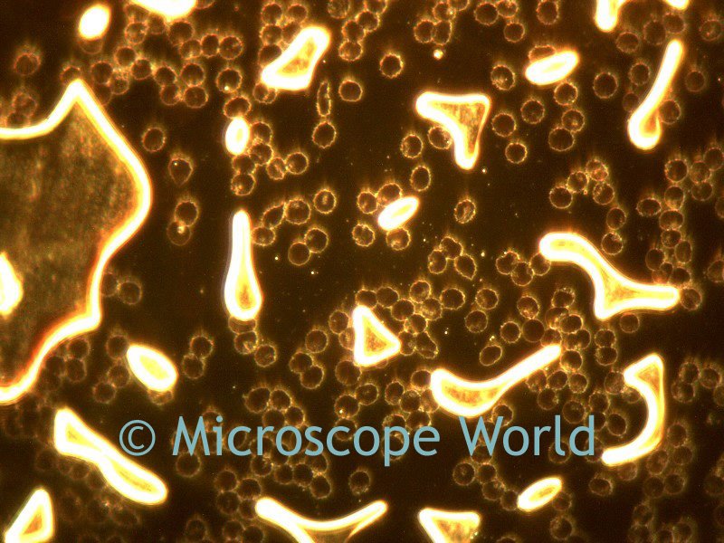
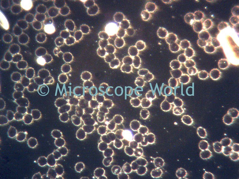
Darkfield is a microscopy technique that is helpful when viewing specimens that have a similar color as their background. Darkfield is used to describe the illumination feature in which the light is dispersed around the sides of the object, in effect giving the specimen a back-lit appearance. It is most frequently used to view live specimen samples that have not been stained.
Darkfield microscopy can be performed with a
stereo dissecting microscope, or a high power
biological microscope.


The above images are live blood cells captured using the
MC2300 microscope digital camera on the
Meiji MT5200 biological microscope with darkfield.

 The above images are live blood cells captured using the MC2300 microscope digital camera on the Meiji MT5200 biological microscope with darkfield.