Wednesday, October 31, 2012

Tips for Cleaning Microscope Stages

Microscope stages and stage plates can get fairly messy from examining biological or industrial specimens. Cleaning these items with a lint free soft cloth and a solution of mild soapy water works best. Don't use an overly wet cloth since moisture may seep into the stand which may have electronics inside. Always unplug equipment before attempting to make repairs or to perform cleaning. Do not use alcohol, acetone or other solvents on your instrument as they may cause damage to painted surfaces.


For glass stage plates, we recommend using mild soapy water and your bare hands. Have a soft paper or cloth towel to set the glass on after you've washed and rinsed your glass.  Carefully dry the glass. It can be a bit damp so let the air dry the glass completely. If you used distilled water, the glass will dry without leaving mineral deposits. Next, by holding the glass by the edges, drop the glass back into the stage and tighten down the locking thumbscrew.

Please Do NOT:
  • Use any aerosol spray product, no matter who sells it or what their claims are.
  • Use eyeglass lens tissue, toilet paper or other paper. It DOES scratch glass!
  • Use pre-packaged cotton balls. Most of the time they are NOT made from cotton.
  • Use lens cleaning solutions marketed by resellers using their brand name. Making your own cleaning solution is cheaper and more effective.

Monday, October 29, 2012

Microscopes and Image Brightness

When using a compound light microscope, there are multiple factors that affect the brightness of your image. We will cover a few of them here.

Numerical Aperture

The numerical aperture (N.A.) is a number that expresses the ability of a lens to resolve fine detail in an object being observed. The higher the N.A. value at a given magnification, the brighter the microscope image will appear.

Magnification

As the magnification of your microscope is increased, brightness of an image is reduced. This is why many times when increasing magnification from 400x to 1000x it becomes important to adjust the iris on the condenser in order to allow more light to reach the specimen.

Beam Splitter

In a trinocular microscope (a microscope with a camera port) the beam splitter is a rod that when engaged, sends more light up toward the camera in order to produce higher quality images. Often when the beam splitter is engaged, one of the eyetubes in the microscope goes dark, or only partial light (maybe 40%) is sent to both eyetubes.

The images below show how as magnification is increased, brightness of microscope images decreases. All images were captured using the MW4-HD2 digital biological microscope.

Duodenum captured at 40x magnification.
Duodenum captured at 100x magnification.
Duodenum captured at 400x magnification.
If you are having trouble getting enough light to your specimen, make sure the condenser is adjusted properly, that the rheostat control on the light is turned up sufficiently, and that the beam splitter is only engaged when capturing images with a microscope camera.

Friday, October 26, 2012

Microscope Eyepiece Diopters

Microscope eyepiece diopters were created to correct the compensation of long- or short-sightedness by adjusting the eyepieces. When you look through a binocular microscope (with two eyepiece lenses), you must be able to change the focus on one eyepiece to compensate for the difference in vision between your two eyes. The diopter adjustment does this.

The microscope eyepiece diopter is adjusted by rotating the top of the eyepiece. Microscope eyepieces with diopters have numbers or marks on them in order to easily adjust eyepiece settings for different users.

How to Properly Adjust Microscope Eyepiece Diopters


The way to correctly adjust a microscope eyepiece diopter is to first close the eye over the eyepiece with the diopter adjustment and normally focus the microscope so that the open eye sees the image in focus. Next, switch eyes (close the open eye and open the closed eye) and without changing the main focus knobs, focus on the image by turning the diopter adjustment only.  Now with both eyes open, the microscope image should be clear with both eyes.  (This technique is also used when using binoculars.)

Wednesday, October 24, 2012

Duodenum under the Microscope

The duodenum is the first section of the small intestine in humans and most vertebrates, including mammals, reptiles, and birds. In mammals the duodenum is thought to be the main location for iron absorption.


Illustration courtesy Olek Remesz

The duodenum precedes the jejunum and ileum and is the shortest part of the small intestine, where most chemical digestion takes place. In humans, the duodenum is a hollow jointed tube about 10–15 inches (25–38cm) long connecting the stomach to the jejunum.

 Cross section of duodenum captured at 100x magnification with the MW4-HD2 digital microscope.

Monday, October 22, 2012

Oil Filter Patch Microscopes

Large machinery saves an enormous amount of time when it is running properly. Downtime however, can waste countless hours, and dollars. Examining the oil filter patch from machinery is a simple process that can result in extending the working hours for machinery and solving problems before they compound and become much more expensive.

Oil filter patches from large machinery.
By using a filter patch microscope, examining particles caught in the oil filter patch is simple. Particles often include teflon, plastic or metal shards. Based on shape, color and tear patterns in the particles found in the filter patch, decisions can be made that will lengthen the life of the machinery.

Metal shaving captured in a filter patch.
Filter patch image captured with the FPHSZ6t3 filter patch microscope system.
When examining oil filter patches typically a top illuminator will suffice. However, having a light that shines up through the filter patch can be helpful in locating debris, especially at higher magnifications. The chart below explains the magnification required to view different particle sizes.


You can view a selection of oil filter patch inspection microscopes here that include both observation microscopes and systems with a camera for capturing images and software for making measurements.

Tuesday, October 16, 2012

Microscope Objectives with an Iris

Why do some objectives have an iris?

In order to preserve darkness of the background for darkfield microscopy, the objective cannot have a numerical aperture (N.A.) higher than the lowest N.A. marked on the darkfield condenser. An iris that can reduce an objective's N.A., can allow you to use higher N.A. objectives for darkfield work. Objectives with an N.A. above 1.2 require an iris for darkfield. For ordinary brightfield observation, the iris can simply stay wide open.

Shown at left is a Plan Semi APO 50x oil objective with an iris. This objective lens has a N.A. of 0.87 and is typically used with the Meiji biological microscopes.

Do I need special objectives for darkfield microscopy?

In most cases, from a transmitted light observation, you will only need a darkfield stop in the condenser. At higher magnifications, you will need an objective with an iris as well as a darkfield condenser.

Friday, October 12, 2012

Immersion Oil or Water Microscope Objective Lens

Should I use immersion oil or water with my microscope objective lens? And why do some objectives require immersion oil or water?


The resolving power of an objective lens depends on its numerical aperture, which in turn depends on the refractive index of the medium between the specimen and the objective lens. A higher refractive index means the lens can gather more light and deliver a better image intensity.

Air has a relatively low refractive index, and when it is the medium between the specimen and the lens, lower N.A. objectives perform at their best capacity. Higher N.A. objectives need a higher refractive index to operate and immersion oil provides that higher index. For optimum performance, you will also need to oil the top lens of the condenser to the bottom of the specimen slide. Immersion objectives are marked "oil" or "oel". Objectives marked "wi", require water as the immersion contact medium.

There are two basic types of immersion oil, Type A and Type B. The only difference between the two is the viscosity. Either will work well. One or two drops of oil are placed on top of the cover slip and the 100x objective lens is brought into position so that it touches the oil and creates a "bridge" of oil between the top of the slide and objective lens. The oil has a refractive index very close to that of glass. This allows very little refraction of the light rays as they go through the slide, specimen, cover slip, oil and through the glass objective lens of your microscope.

Wednesday, October 10, 2012

Stereo Fluorescence Microscopy

Stereo Fluorescence

What exactly is Fluorescent Microscopy?

A fluorescence microscope uses fluorescence and phosphorescence instead of (or sometimes in addition to) reflection and absorption to study properties of an organic or inorganic substance. The stereo fluorescence microscope illuminates a sample with light of a wavelength which excites fluorescence in the sample. The fluoresced light, which is usually a longer wavelength than the illumination, is then imaged through a microscope objective and two filters are used - the illumination (excitation) filter which ensures the illumination is near monochromatic and at the correct wavelength, and a second emission filter which ensures none of the excitation light source reaches the detector.

How does Fluorescent Microscopy Work?

In most cases, the sample of interest is labeled with a fluorescent substance called a fluorophore and then illuminated through the lens with the higher energy source. The illumination light is absorbed by the fluorophores (now attached to the sample) and causes them to emit a longer, lower energy wavelength light. This fluorescent light is then separated from the surrounding radiation with filters designed for that specific wavelength, allowing the viewer to see only that which is fluorescing.

The basic task of the fluorescence microscope is to let excitation light radiate the specimen and then sort out the much weaker emitted light from the image. First, the microscope has a filter that only lets through radiation with the specific wavelength that matches your fluorescing material. The radiation collides with the atoms in your specimen and electrons are excited to a higher energy level. When they relax to a lower level, they emit light. To become detectable (visible to the human eye), the fluorescence emitted from the sample is separated from the much brighter excitation light in a second filter. This works because the emitted light is of lower energy and has a longer wavelength than the light that is used for illumination.

Stereo Fluorescent Microscope

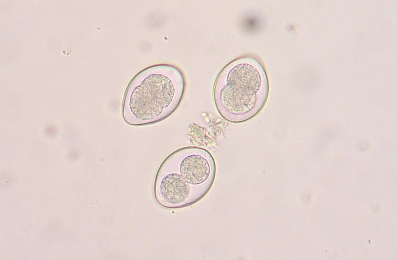
Whether working with C. elegans, drosophila or zebrafish, developmental biology depends on stereo fluorescence to sort eggs or embyros that have been tagged with a specific marker called a fluorophore. Typically stereo fluorescence microscopes are expensive and therefore shared within a lab or facility where time is rigidly scheduled and many users can be accommodated.

Microscope World has developed a less expensive alternative which requires no compromise in quality and allows use of an existing stereo microscope for stereo fluorescence. Many stereo microscope systems can be adapted into a stereo fluorescence microscope, resulting in quite a cost savings, but sacrificing no quality when it comes to fluorescence.

Additionally, the new SXT500 comes available from Microscope World either as a complete system, including a high quality common main objective stereo microscope system, or as an illumination system you can use with many models of dissecting microscopes. If you are interested in getting information on adapting your existing stereo microscope for stereo fluorescence contact us.

Monday, October 8, 2012

Student Microscopy Tips

There are a few tips that when followed, will make student microscope work a little less frustrating. Here are our top ten tips for a good microscopy experience:

  1. A compound microscope will give a two-dimensional, flat image. Use a compound microscope to examine specimens on prepared slides. Compound microscopes are available as either monocular (1 eyepiece), binocular (2 eyepieces) or trinocular (2 eyepieces + camera port).
  2. A stereo microscope has a binocular body and will provide a three-dimensional image. Use a stereo microscope for specimens that have depth or are large in size and require greater working distance. Stereo microscopes are perfect for items you can hold and view in your hand, but wish to view more magnification (flowers, a dollar bill, rocks, etc.)
  3. Start to focus using the lowest magnification. In a compound microscope, start with the 4x objective. Make sure the objective is clicked into place. With a stereo microscope, start by using the 1x objective. Once the specimen is in focus at a lower magnification, increase magnification.
  4. Always place the specimen in the center of the stage or stage plate. When using a compound microscope make certain the slide is placed on the stage with the light centered over it.
  5. When viewing a slide, make sure it is the right side up. This is especially important when using a prepared slide. If upside down, it will not be in focus at high power.
  6. Focus first using the coarse adjustment, then use the fine focus. You should be able to change from one objective (magnification) to another with just a minor fine focus adjustment. If you can do this, it means your microscope is parfocal.
  7. Adjust the illumination by using the intensity control and condenser or diaphragm.
  8. Remember when using higher magnifications you will need to adjust the light source.
  9. Always use proper care and handling of the microscope. Carry it with two hands, one hand around the arm of the microscope and the other hand under the base.
  10. Keep your microscope clean. To clean the lenses, first remove any dust and dirty by using a camel hair brush or canned air. Moisten the end of a Q-tip with lens cleaning solution. Keep the other end dry. Clean the optical surface with the moist end of the Q-tip using a circular motion. A solution of Windex with vinegar will also work. Use a dust cover when the microscope is not being used. Learn more microscope cleaning tips here.

Wednesday, October 3, 2012

Parasites in Sheep & Goats

Two of the largest problems goat farmers face are worm and coccidia infestations. These two parasites alone kill more goats than all other illnesses combined. Surprisingly, many goat farmers do not have an established program of regular, systematic microscopic examination of goat feces for these parasites. Analyzing the feces for these parasites is a simple process and can easily be incorporated into the overall goat health program.

Coccidia is a protozoan parasite. Coccidiosis, that malady caused by Coccidia, can be one of the most economically devastating diseases in many livestock. It can be especially harmful to recently weaned kids. It causes a watery diarrhea that is sometimes bloody and can even be a life-threatening problem to an especially young animal. The presence of Coccidia in the intestines of an individual does not mean the animal is actually suffering from coccidiosis as Coccidia are found everywhere. These protozoans only cause disease when their numbers become so great that damage is done to the host.

Some species of Coccidia found in animals:
  • Eimeria - goats, swine, horses, cattle, sheep, poultry, rabbits
  • Toxoplasma - cat family only
  • Isospora - dogs, cats, primates, swine
  • Neospora Caninum - dogs
  • Sarcocystis - carnivores and omnivores
  • Cryptosporidium - broad host spectrum including humans
Below is an image of a highly magnified view of Coccidia Sporogony. This is the infective stage of the Coccidia when it is consumed by the animal. Species of Coccidia are determined by looking at the structure of this stage. The oocyst contains two sporocysts, and this is typical of the genus Isospora (as well as Toxoplasma, although Toxoplasma oocysts are much smaller). Sporulated oocysts of the genus Eimeria contain four sporocysts. The oocysts are typically between 35-50um long. At 100x magnification about 40 of them would appear in the field of view. Although they appear small, you can see and identify them.

Goat oocyst.
In order to examine your animals for worms, eggs and coccidia, you will need a microscope and some basic supplies. The microscope does not need to be advanced - a simple high school model such as the MW2-H3 microscope will allow you to view eggs, worms and coccidia. You will need blank glass microscope slides, cover slips, cheesecloth or a strainer, test tubes, a stirring rod (use a chopstick!), fecal flotation solution (sugar or salt works) and a test tube holding rack. If you do not want to purchase a rack, simply punch holes in a cardboard box or styrofoam block to hold the tubes vertically.

FECAL ANALYSIS:

  1. Mix up the flotation solution.  It should be saturated.  This means that you dissolve as much solid in the water as it will hold.  You can use a variety of chemicals including salt or sugar. Saturated sugar is prepared by dissolving a pound (454 grams) of sugar in 1 1/2 cups (355 ml) of water, and saturated salt takes a pound (454 grams) of salt in 4 4/5 cups (1140ml ) of water.   If there are salts left in the bottom of the liquid, pour off the saturated liquid into a new container.
  2. Collect fresh feces.  Use an old pill bottle or a small jar for each animal.  Be sure to label the container with the date, time and animal that provided the specimen.
  3. Place 3 or 4 fresh goat pellets (one to three grams) into a test tube and pour in just enough flotation solution to cover them.
  4. Mash them up in the liquid with your stirring rod.  Add more of the solution and pour it through the strainer or cheesecloth to remove the large particles.  Pour the strained liquid into a clean test tube.
  5. Fill up the test tube to the very top with more liquid.  Place a microscope coverslip over the top.  There should be no air between the coverslip and the liquid. Over time (20-30 minutes) the eggs will float up to the top and adhere to the glass plate.
  6. Carefully remove the coverslip and lower it at an angle over a microscope slide with the sample sandwiched between both pieces of glass.
  7. Examine the specimen for worm eggs and coccidia oocysts.  Start with the lowest power (40x) on your microscope and carefully move up to 100x  and even 400x if you see something interesting.  An illustrated chart would be helpful in identifying the parasites.  Note, you will also be looking at other debris.  Do not confuse it with parasites.
  8. You should be able to see coccidia oocysts, nematode eggs, and some tapeworm eggs.  Nematode eggs are shed by a large number of nematodes (worms), most of which cannot be easily distinguished from each other with this type of procedure.  This group is referred to as strongyle eggs and worming recommendations can be based on the quantity of strongyle eggs present.  Since fecal counts only estimate the parasite load, there is no clear cut level when worming should be undertaken.  As a general guide, a level of about 500 eggs per gram of feces would indicate that worming is required for sheep, goats, or cattle. A better way of deciding when to treat would be to monitor fecal samples every 4-8 weeks and worm when there is a dramatic rise in egg counts.
Remember, there are different treatments for various parasites. In some cases changing the environment (a new pasture), may be all that is required. When worming medications are used, be sure to use one that is effective against the parasites for which you are treating. It is counterproductive to treat for everything, when you only need to treat a specific parasite. If unsure of the type of infestation, consult your local veterinarian.

Coccidia oocysts in a fecal flotation from a cat. Image courtesy Joel Mills.

Monday, October 1, 2012

Increasing Microscope Magnification

Increasing microscope magnification can be a delicate balance between an increase in magnification and a loss in resolution. Before we discuss increased magnification, it is good to make sure it is understood where total magnification comes from. Microscope magnification is a combination of the eyepiece magnification and the objective lens magnification.

In the stereo microscope image above you can see the eyepieces (which are changeable) and the objective lens (which is built into the microscope and therefore not changeable). On a standard stereo microscope (not a common main objective stereo microscope) the objective lens is built into the microscope and the only way to change this magnification is by adding an auxiliary lens to the existing objective lens. These are typically available in increments of 0.5x, 0.75x and 1.5x magnification. The most common way to increase magnification on a stereo microscope is by adding one of these auxiliary lenses. Keep in mind however, that with added magnification comes decreased field of view. Another common problem that accompanies increased magnification on a stereo microscope is the increase need for brighter illumination. Typically anything above 80x magnification on a stereo microscope requires the minimum of a 150w halogen fiber optic ring light or fiber optic pipe illuminator.

In the biological microscope image above note the eyepieces and the objective lenses. These provide total magnification for the microscope. Both can be changed. However, many times the microscope user thinks the easiest way to increase magnification is by simply increasing the magnification of the eyepieces. To maintain useful magnification with satisfactory clarity and resolution, it is important to avoid empty magnification or making the specimen appear bigger but not clearer. In general, total magnification should not exceed 750x-1000x the N.A. of the objective. For example, with a 40x, N.A. 0.65 objective, the total magnification should be between 480x and 650x. Using this example, if you were to try to use 20x eyepieces with this 40x objective lens, you would end up with 800x magnification - a number that is outside this range. Pairing these higher magnification eyepieces with the 40x objective would result in an image that is in fact magnified, but does not have clear and crisp resolution.