Trachea, 100x magnification.
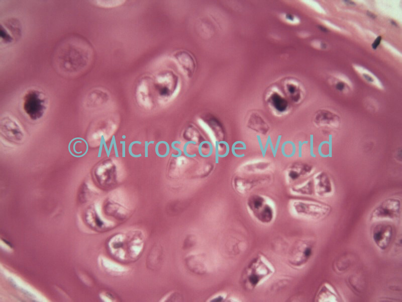 Trachea, 400x magnification.Trachea, 100x magnification.Trachea, 400x magnification.
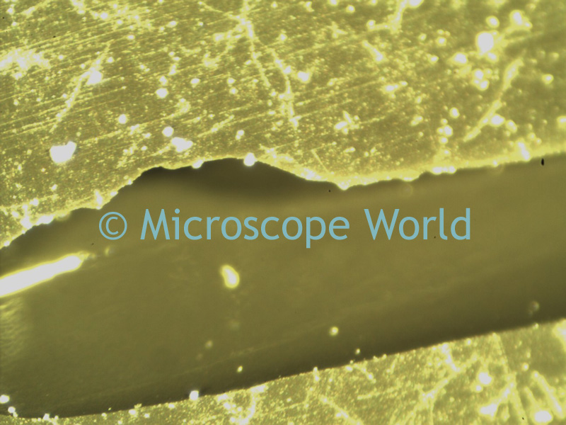 The two images above were captured using the MC2000 microscope digital camera on a National Optical 420T stereo microscope. These images were captured at 30x magnification. In order to view scratches or minor flaws in metal, a stereo microscope works well.
 In more advanced instances, when more magnification is required a metallurgical microscope is best. The image above was captured with the Meiji MT7100 metallurgical microscope at 200x magnification. The metallurgical microscope is similar to a biological microscope, in that it has high power objectives. On a metallurgical microscope however, the light shines down from inside the objective directly onto your specimen, allowing you to view opaque samples at high magnification.
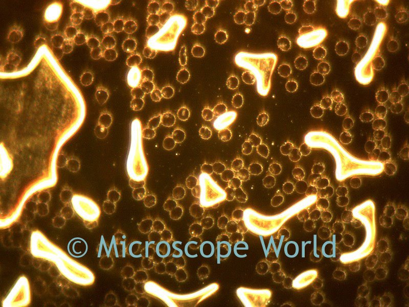
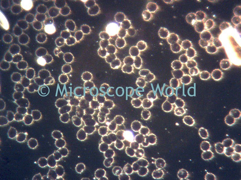 The above images are live blood cells captured using the MC2300 microscope digital camera on the Meiji MT5200 biological microscope with darkfield.
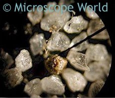
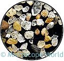 The colors of sand vary greatly depending on the area the sand came from. Some sand is created from river rocks that eroded from riverbanks. Other particles of sand look like small polished pieces of rock. There is even a beach in California called glass beach! This beach was created many years ago when residents used to dump garbage near the coast of a town near Fort Bragg, California. Years of waves crashing on the beach have polished small pieces of glass into colorful pebbles!
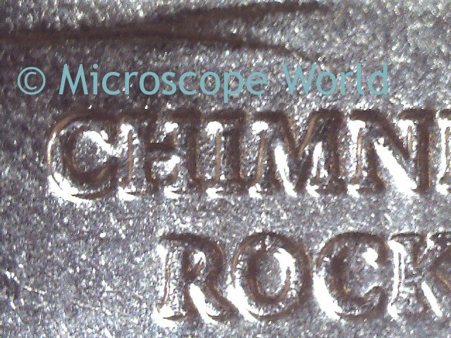
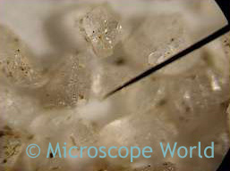 You can view more images of sand under a microscope here.
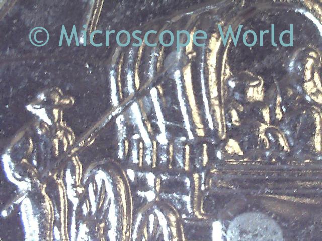 When viewing coins a magnification of 10x - 30x is usually ideal.
 The above image is human smooth muscle under a microscope at 400x magnification. This image was captured with a Canon SLR digital camera using the CSLR microscope camera adapter on the 163 compound microscope.
 A microscope c-mount adapter allows you to connect a microscope camera to the trinocular port of your microscope. C-Mount adapters are microscope-specific, which means that if you are using a Meiji microscope, you need the Meiji c-mount adapter to connect your camera. Image at left is a Motic C-Mount adapter for the SMZ stereo microscope.
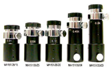 Meiji C-Mount adapters varying from 2.5x - 0.3x.
 Image courtesy of Martin Moe.
 This image of foam was captured with the PRCFscan microscope camera. This camera is exceptional for research applications or tasks where light is low. Generally when working in low-light conditions, a camera with a CCD chip is best for obtaining a crisp and clear resolution image. Cameras with CMOS chips are better for routine image capture and everyday use. CMOS cameras are often used in industrial settings where extreme detail in the photograph is not as important.
 Microscope World is excited to announce the launch of its blog. In this area you will find information on microscopes, microscopy and all-things-microscope. We regularly capture images with the microscope and hope to be able to share these with you as well.