Monday, November 30, 2009

Microscope Project: Mold

Next time you open the fridge and see a moldy piece of bread, cheese or a strawberry, don't throw it out right away. Grab your microscope and learn a little bit first! If you have both a stereo microscope and a compound microscope, you can use each to view the mold.

Starting with the stereo microscope, take a look at the mold. Notice the texture of the mold, it may almost look soft like fabric.

Image of moldy bread at 20x magnification courtesy of King Edward VII School.

Now take the same moldy food and scrape a small piece of mold off with a knife. Place the mold on a blank microscope slide and place a cover slip on top of it. Using a high power microscope, take a look at the mold again. How does it look different? Can you identify any bacteria? Draw a picture of the images that you see and ask your science teacher to help you identify the cells in the mold.

Friday, November 27, 2009

Identifying Particulate Contaminates

Microscope World often helps customers configure a microscope system for identifying particulate contaminates. For example, the standards for European Pharmacopoeia Section 2.9.19, Method II: Microscopic Particle Count Test require the following:
  1. Binocular Microscope
  2. Eyepiece Reticle (Graticule) for specified type
  3. Stage Micrometer with Certification
  4. Mechanical Stage
  5. Episcopic (Reflected) Light Microscope
  6. External Illuminator with Oblique Lighting
  7. 10x Eyepieces with a 10x Objective Lens
The following microscope system was created to meet these standards. The MT7200 reflected light metallurgical microscope, paired with an IMA/USP counting reticle, stage micrometer (NIST certified), and a LED dual pipe illuminator for oblique lighting.

One of our microscope specialists configured this microscope system to meet specific standards.

If you have the need to identify particulate contaminates please give us a call. We would be happy to put a custom quote together to meet your needs.

Tuesday, November 24, 2009

What Can I See With My Microscope?

A compound light microscope has a resolution of about 200nm. A light microscope will allow you to view cells that are alive, fixed or stained.

Bacteria (shown above) is an example of one of the smallest cells that can be seen with a light microscope. Animal and plant cells can be viewed with a light microscope as well.

In order to view small molecules or viruses, a transmission electron or scanning electron microscope is needed. These types of microscopes have a limit of resolution down to 2nm.

Monday, November 23, 2009

Tropism

Tropism is the biological phenomenon that indicates growth or movement of a biological organism (usually a plant) in response to an environmental stimulus.

Photo courtesy of Mark Simmons.

The above tropism is a polarized light microscope image of adipic acid, (CH2)4(COOH)2. The nearly 2.5 billion kg of adipic acid produced each year is mostly used as a monomer for the production of nylon. Other major uses involve polymers. It is a monomer for production of Polyurethane and it is used in making PVC. The image above was captured using a microscope digital camera adapter and a standard digital camera.

Wednesday, November 18, 2009

Microscope Sale

Meiji microscope closeout special. While supplies last, this Meiji microscope system, including fluorescent ring light is $699. High quality Japanese optics. Your choice of 10x, 20x, 30x or 40x magnification. Order online here.

Tuesday, November 17, 2009

Stereo Zoom Microscopes

The Meiji EMZ-13TR stereo microscope was used to capture these images of capacitors.

This image was captured at 75x magnification. In order to achieve this magnification, 20x eyepieces, a 1.5x auxiliary lens and a 1.0x c-mount adapter were used.

This image was captured at 150x magnification.

Monday, November 16, 2009

Hair Science Project

The goal of this science project is to measure hair damage and strength using no hair products and then again after using a variety of different products.

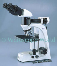
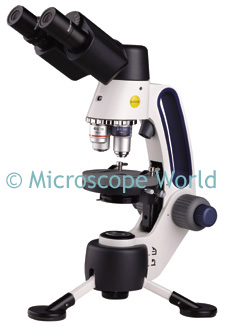
You will need a student microscope with at least 100x magnification. When viewing a single strand of hair, the biological elementary student microscope works well.

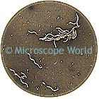
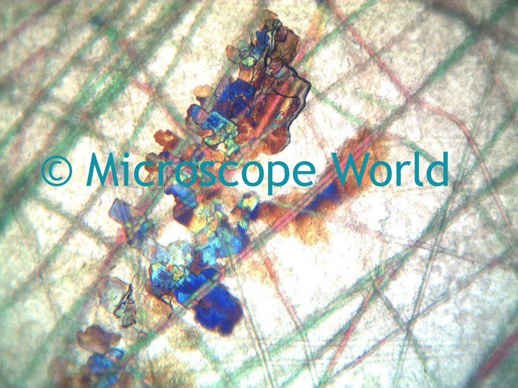
Before you begin, find a few friends or family members willing to participate in your science experiment. It is best to use hair that has not been lightened or darkened, as this alters the strength of the hair. Once you have located your subjects, gather a clean strand of hair from each and place it under the microscope. If you can, capture an image of each and label the image with the name, magnification and test # (see example below).

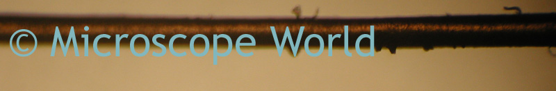
Friend A, 100x magnification, test #1

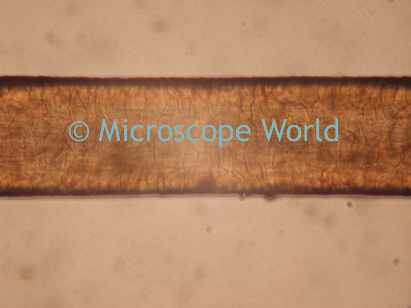
Friend A, 1000x magnification, test #1

After capturing your images, set the pieces of hair aside in a labeled envelope. Later you will want to pull these out again to test the strength of the hair.

Once you capture the images, have each friend use the same hair care product for a week. Try something that advertises "hair strengthening" or "heals damaged hair". After a week, collect a piece of hair from each person and look at it under the microscope.

Do you notice any difference between the hair from test #1 and test #2? Does the hair care product really make the hair healthier? A damaged piece of hair will have rough edges when viewing it through the microscope. You may want to have the subjects try another week of the hair product and compare a third time.

Finally, take the hair from each session and try to break it by pulling on each end. Do you notice a difference in the strength of the hair that was not treated with any products to those that were?

Did you notice a specific color of hair tended to be stronger than others? If so, you could complete a secondary science project by gathering more samples of similar colors of hair for comparison. If you have great findings from your science project, send us an email, we would love to hear about it!

Friday, November 13, 2009

Nature at 10x and 30x Magnification

Stereo or dissecting microscopes allow you to view nature up close and in detail. Even a simple 20x kids microscope can reveal the stigma of a flower or plant. When viewing items found in nature, a microscope with 10x - 40x magnification is generally best.

This leaf image was captured at 10x magnification,

The same leaf, at 30x magnification.

If you are looking for a good children's microscope that connects to the computer so you can capture and save your nature images, Microscope World recommends the DS2 digital student microscope.

Friday, November 6, 2009

Coin Collecting Microscopes

Coin collecting requires good optics and low magnification in order to fit the entire coin in the field of view. You can view a variety of coin collecting microscopes here.

These images were all captured with the SMZ-168 stereo microscope with 5x eyepieces. The MC1000 microscope camera was mounted over the 5x eyepiece in order to have a lower magnification. This same result could also be obtained using the 10x eyepieces and a 0.5x auxiliary lens.

Tuesday, November 3, 2009

Swift Microscopes

The full line of Swift Microscopes can be found at Swift-MicroscopeWorld. In February of 2010 the micro/macro M3 microscope is being introduced. This microscope allows students to view both micro and macro specimens. The macro setting (20x magnification) is perfect for viewing rocks, plants and insects. The micro magnifications of 40x, 100x and 400x provide standard biological magnifications and allow the student to view protozoans, blood cells, etc.


Pre-orders are being accepted for the Swift M3 microscope, due to ship February 2010.