
 Captured at 100x magnification, this image is a bone marrow smear from a patient with chronic myelogenous leukemia.
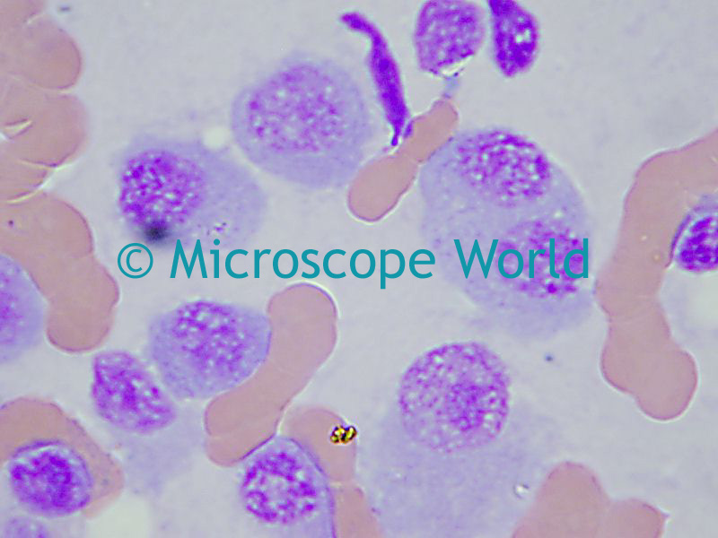 Captured at 1000x magnification, this image is a bone marrow smear of a myeloma case. You may noticed the binucleated plasmacyte, partly covered by the word "World".
These images were captured using a biological laboratory microscope.