Friday, January 29, 2016

Cirrhosis of the Liver

The liver weighs about three pounds and is the largest solid organ in the body. The liver transports oxygen, manufactures blood proteins that aid in clotting, and aids in the body's immune system function. The liver stores excess nutrients and returns them back to the blood stream.

The liver produces bile required for food digestion and helps the body store sugar in the form of glycogen. It breaks down saturated fats and produces cholesterol. Harmful substances in the body such as alcohol or drugs are removed from the body by the liver.

Cirrhosis is a disease in which healthy liver tissue is replaced slowly with scar tissue, eventually damaging the liver from functioning properly. The scar tissue blocks the blood from properly flowing through the liver, and slows the process of nutrients, hormones, toxins, drugs and naturally produced toxins. Cirrhosis also slows the production of proteins and other substances made by the liver.

According to the National Institutes of Health, Cirrhosis of the liver is the twelfth leading cause of death by disease.

Hepatitis C, fatty liver and alcohol abuse are the most common causes of cirrhosis in the liver in the United States but anything that damages the liver can cause cirrhosis, such as obesity and Diabetes (fatty liver), viral infections of the liver, repeated heart attacks and certain inherited diseases.

The images below of Cirrhosis of the liver were captured using the RB30 biological lab microscope and a microscopy USB camera at Microscope World.

Microscope World image of cirrhosis of the liver at 40x magnification.
Cirrhosis of the liver under the microscope at 40x.

Cirrhosis of the liver image captured at 100x magnification at Microscope World.
Cirrhosis of the liver under the microscope at 100x.

Cirrhosis of the liver captured at 400x by Microscope World.
Cirrhosis of the liver under the microscope at 400x.

Microscope World image of cirrhosis of the liver at 400x.
Cirrhosis of the liver under the microscope at 400x using a Plan Fluor Objective.

Wednesday, January 27, 2016

Quality Control Microscopes

There are a variety of quality control microscope options and depending on your specific needs, this post should help sort out the options.

Basic Inspection of Small Parts

Quality control microscopes with zoom magnification.
S6-ILST Zoom Microscope
For basic inspection of small parts a sturdy quality control stereo microscope will meet all needs. Depending on your budget, there are two options:
  1. A dual power stereo quality control microscope.
  2. A stereo zoom quality control microscope.
With a dual power stereo microscope you will view two set magnifications only. These microscopes are less expensive and the most commonly purchased microscope have 10x / 30x magnification. At 30x your field of view is about 15mm and you would be able to identify flaws that are 1-2mm in size. If you are looking at smaller samples or defects a 15x / 45x microscope is a good alternative.

A stereo zoom quality control microscope is going to provide a lot more control over the magnification. A good magnification range is 7x-45x. This range can be increased even further with higher magnification eyepieces or an auxiliary lens. This magnification chart shows some of the magnification range options of a stereo zoom microscope when different eyepieces or auxiliary lenses are used.

Larger Sample Inspection

Microscope World Articulated Arm Stereo Zoom Microscope System
Articulated Arm Stereo Microscope
When viewing larger samples a stereo boom microscope will provide extra working space beneath the microscope. These microscopes typically use either a dual pipe light or an LED ring light to flood the working area with light. Again, the magnification range of 7x-45x is typically plenty for quality control inspection.

There are several types of boom stands used in industrial settings. The articulated arm microscope (shown at left) provides a lot of options for exact positioning of the microscope body above the sample. Another type of inspection microscope for large parts is a standard boom stand microscope, or a ball bearing boom stand microscope. The ball bearing boom stand microscope is used when the head of the microscope needs to be slid out of the way frequently.

Handheld Inspection Microscopes


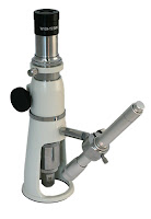
Handheld inspection microscope
There are several options for handheld inspection microscopes. The most common handheld inspection microscope is also known as a shop microscope (shown at left). This microscope has a single magnification and a built-in eyepiece reticle for making measurements. The handheld inspection microscope is placed directly on top of the object that needs to be viewed and when looking through the eyepiece the ruler is imposed directly on the sample, making it easy to quickly make measurements.

Digital Inspection Microscopes

Digital Inspection Microscope for Quality Control

The other type of quality control inspection microscope is a digital inspection microscope. This microscope does not have any eyepieces to look through, instead all images are displayed directly on a monitor or a computer. The included software allows image capture, making measurements and adding annotations to images. The digital handheld inspection microscope is perfect for documenting quality control issues and emailing images for co-worker inspection.


If you have a specific quality control microscope system or process you are trying to meet, contact Microscope World for a solution.

Wednesday, January 20, 2016

Prostate Cancer under the Microscope

Carcinoma of the Prostate, also known as Prostate Cancer, is the development of cancer in the prostsate. The prostate is a walnut sized gland in the male reproductive system. Most prostate cancers grow slowly, but the cancer cells may spread to other parts of the body, in particular the bones and lymph nodes.

Late stages of prostate cancer can cause symptoms such as difficulty urinating, blood in the urine, or pelvis or back pain while urinating. Feeling tired is another common symptom as the cancer causes low levels of red blood cells.

Prostate cancer is diagnosed by a biopsy. The images below of prostate cancer were captured using the RB30 biological lab microscope and the DCM5 microscope camera (3 megapixels).

Microscopy image of prostate cancer at 40x.
Prostate cancer under the microscope at 40x magnification.

About ninety-nine percent of prostate cancer cases occur in men over the age of fifty. Diets high in processed meat, red meat or milk products and low in vegetables can increase prostate cancer risk. 

Lab microscope image of prostate cancer at 40x from MicroscopeWorld.com.
Prostate cancer under the microscope at 100x magnification.

Prostate cancer screening is controversial because prostate-specific antigen (PSA) testing increases cancer detection but does not decrease mortality. PSA testing sometimes results in over-diagnosis and over-treatment as most cancers diagnosed would remain asymptomatic.

Prostate cancer image from MicroscopeWorld.com captured at 400x.
Prostate cancer under the microscope at 400x magnification.

The five-year survival rate for prostate cancer in the United States is ninety-nine percent.

Microscope World image of prostate cancer at 400x.
Prostate cancer under the microscope at 400x magnification using Plan Fluor objective lens.

You can learn more about prostate cancer here.

Wednesday, January 13, 2016

Student Project: Soil Under the Microscope

Soil contains a variety of particles, bacteria or even living creatures and can be viewed with a compound biological microscope or with a stereo dissecting microscope.  When using a compound biological microscope to view soil you will want to place only a few particles of soil onto a microscope slide and put a cover slip on top to flatten out the sample. Start at the lowest magnification and work your way up from 40x, to 100x and then finally 400x. Can you identify any living organisms? If you have a camera, capture the image and note the differences between the lowest and highest magnification. If you don't have a camera just sketch what you see under the microscope. Were you able to find any living organisms or maybe a small worm?

If you are using a stereo dissecting microscope, placing a bit more soil on the glass stage plate won't be a problem, and you do not need to cover it with anything since the stereo microscope has a greater working distance.

The images of soil shown below were captured with the UX1 High School Compound Microscope using the DCM5 microscope camera with 5 megapixels. Notice some of the cracks in the dry dirt in the first image. And in the final image captured at 400x can you see some of the green moss?

Soil under the microscope at 40x.
Soil captured under the UX1 High School Microscope with the DCM5 camera at 40x.

Dirt captured under the microscope at 100x magnification.
Soil captured under the UX1 High School Microscope with the DCM5 camera at 100x.

Soil under the microscope at 400x magnification.
Soil captured under the UX1 High School Microscope with the DCM5 camera at 400x.

Monday, January 11, 2016

Viewing Bacteria with a Microscope

Bacteria can be hard to identify on an unstained microscope slide. Bacteria are difficult to identify under a brightfield microscope because they lack color, are small and also transparent. In other words, bacteria often look similar to the background they are floating around in. Amateur microscopists may also have trouble distinguishing between bacteria and dust or debris in the sample.

Phase contrast microscope image of bacteria.
Bacteria captured under a phase contrast microscope.

The two solutions for the tricky problem of viewing bacteria involve one of the following:

  1. Staining the microscope slide.
  2. Using a phase contrast microscope.

Microscope cell staining is a technique used to enable better visualization of cells and cell parts under the microscope. By using different stains, bacteria or a cell wall are easier to identify. Most stains can be used on non-living (fixed) cells, while only some types of stain can be used on living cells. There is an extensive list of the types of stains and those that can be used on living cells here.

Phase Contrast Microscope from Microscope World
Phase Contrast Microscope

Phase contrast microscopes allow researchers to observe differences between structures that have a similar level of transparency. By eliminating the need for time-consuming staining, phase microscopes allow research to be conducted more quickly and efficiently. A phase contrast microscope is different from a brightfield microscope in three ways. The phase contrast microscope utilizes a phase contrast condenser, phase contrast objective lenses and a phase centering telescope to center the condenser. You can learn more about phase contrast and centering the phase contrast condenser here.

Tuesday, January 5, 2016

Villous Adenoma under the Microscope

Villous adenoma is a type of polyp that grows in the colon, anywhere else in the gastrointestinal tract, or occasionally other parts of the body. These polyps start out benign, but can become malignant. Villous adenoma has been demonstrated to contain malignant parts in up to one third of affected persons and invasive malignancy in another one third of removed samples. If the affected polyp is large it may be necessary to remove part of the colon.

Villous adenomas can cause severe diarrhea. Since Villous adenoma often create mucus, it can cause the infected perfson to get hypokalemia, which means they have a low level of potassium in the blood.

The images below of Villous adenoma were captured using the RB30 biological microscope and the high definition HDCAM7 microscopy camera.

Colon polyp image under the microscope at 40x.
Villous Adenoma under a biological lab microscope at 40x.

Villous Adenoma under the microscope at 100x.
Villous Adenoma under a biological lab microscope at 100x.

Colon polyp under the microscope at 400x.
Villous Adenoma under a biological lab microscope at 400x.

Villous Adenoma under a biological lab microscope using a Plan Fluor Apochromat 40x objective.