 |
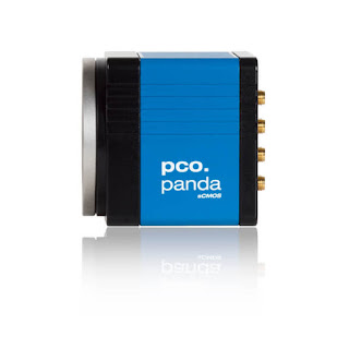
| pco.panda 4.2 Camera |
The pco.panda 4.2 camera combines revolutionary sCMOS technology in a compact design. High quantum efficiency (up to 80%) and low readout noise make this camera suitable for countless applications. The USB 3.1 interface enables ultra-speed data transfer and direct power via the USB cable, making external power supplies redundant. Frame rate = 40 fps @ full resolution 2048 x 2048, 80 fps @ 2048 x1024.
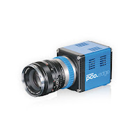
The pco.edge 3.1 camera has a sCMOS sensor and is designed for users who require high resolution and high frame rates. This USB3 camera is available in color or monochrome. 50 fps @ full resolution 2048 x 1536, 75 fps @ 1280 x 1024, 160 fps @ 640 x 480.
The pco.edge 4.2 camera has sCMOS technology and can be optionally upgraded with a water cooling system. This camera has high quantum efficiency at up to 82% at peak. 100 fps @ RS fast scan 2048 x 2048, 189 fps @ fast scan 1920 x 1080, 420 fps @ fast scan 640 x 480 when using camera link.
The pco.edge 4.2 LT camera has sCMOS technology and USB3 output. High quantum efficiency of up to 82% at peak. 40 fps @ full resolution 2048 x 2048, 80 fps @ 1280 x 1024, and 170 fps @ 640 x 480.
The pco.edge 5.5 camera has sCMOS technology and can be optionally upgraded with a water cooling system. Quantum efficiency of > 60% @ peak. With camera link, the frame rates are high: 100 fps @ RS/GR fast scan 2560 x 2160, 201 fps @ fast scan 1920 x 1080, 450 fps @ fast scan 640 x 480.


