Wednesday, November 29, 2017

Fluorescence Microscopy

Fluorescence microscopy is an essential tool in biological and biomedical sciences, as well as in material science. The fluorescence images below were captured using a research microscope with a research microscopy camera.

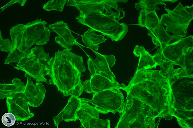
Green fluorescence microscopy image
Green fluorescence image captured with a research microscope.

Monochrome fluorescence microscopy image
Monochrome fluorescence image captured with a research microscope.

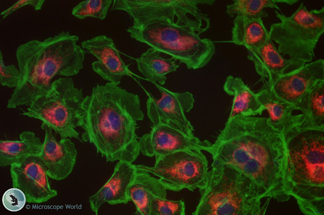
Fluorescnece microscopy image showing multi channel fluorescence.
Multi channel fluorescence microscopy image captured with a research microscopy camera.

For more information on fluorescence research microscopes or research microscopy cameras for your specific application contact Microscope World.

Thursday, November 9, 2017

Tissue Biopsy under the Microscope

A biopsy is a way for a doctor to diagnose a disease. A sample of tissue or cells is removed from the patient by a doctor and sent to a pathologist to be examined under the microscope.

The following images were captured under a pathology microscope during a tissue biopsy.

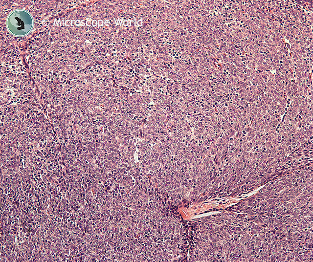
Microscopy image of tissue biopsy.
Tissue biopsy captured under a pathology microscope.

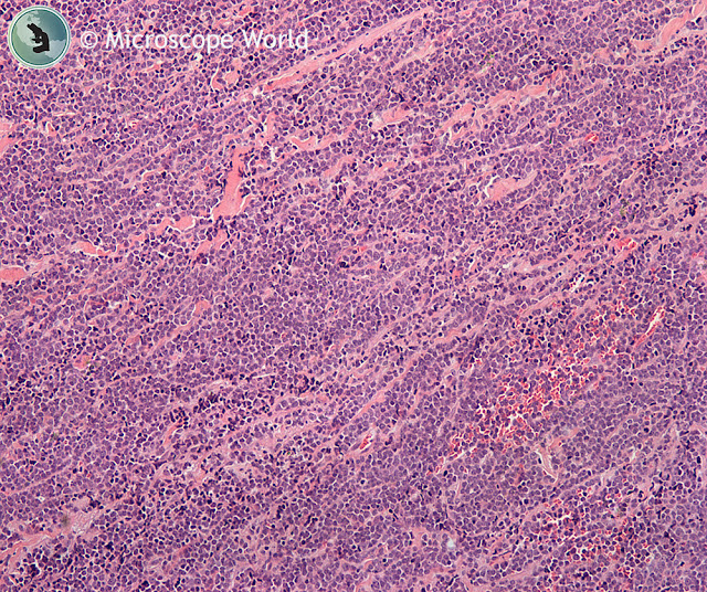
Microscopy image of tissue captured under a pathologist microscope.
Tissue biopsy captured under a pathology microscope.

Tissue biopsy captured under a pathologist's microscope.
Tissue biopsy captured under a pathology microscope.

Tissue biopsy captured under a pathologist's microscope.
Tissue biopsy captured under a pathology microscope.

To learn more about different types of biopsies click here. For more information on pathology microscopes contact Microscope World.