Wednesday, June 28, 2017

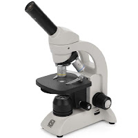
Microscope Ergonomic Work Station

A proper ergonomic work station when using your microscope can reduce the risk of occupational injuries. UCLA Ergonomics suggests the following tips to maintain a safer environment when using microscopes:
  • Sit in the proper position keeping your back straight and your shoulders back. Distribute your body weight evenly on both hips, and keep your feet flat on the floor.
  • Arrange your work space so that it is close to you.
  • Ensure there is proper padding if leaning on hard surfaces.
  • Work with elbows in close proximity to the body.
  • Work with wrists in a straight and neutral position.
  • Adjust and/or elevate your chair, workbench, or microscope as needed to maintain an upright head position.
  • Adjust microscope eyepieces or mount the microscope on a 30° stand for easier viewing.
  • Repair and clean microscopes regularly. Learn how to clean lenses here.
  • If possible, spread microscope work throughout the day and between several people. 
  • Schedule works breaks. Every 15 minutes, close your eyes or focus on something in the distance. Every 30 minutes get up to stretch and move.
Microscopy is a demanding and rewarding field to work in. By maintaining an ergonomic work space you can ensure years of healthy and pain-free work.

Sources:
"Tips for Microscopy." Laboratory Ergonomics. Ergonomics.ucla.edu, 2012. Web. 27 June 2017.
"Posture for a Healthy Back." Articles. Health. Clevelandclinic.org, 2017. Web. 27 June 2017.
Wednesday, June 21, 2017

Persimmon Endosperm

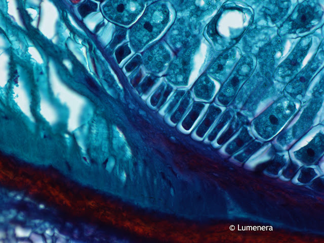
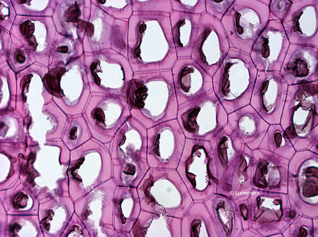
Endosperm is a tissue produced inside seeds following fertilization in most flowering plants. It surrounds the embryo and provides nutrition in the form of starch, though it can also contain oils and protein. This can make endosperm a good source of nutrition in the human diet. The image below is of a persimmon endosperm captured under the microscope using the Lumenera Infinty 2-1 color microscopy camera.

Persimmon endosperm captured under the microscope with Infinity 2-1 microscopy camera.
Persimmon Endosperm under the Microscope captured with Infinity 2-1 Camera

Contact Microscope World with questions regarding microscopes or microscopy cameras.

Wednesday, June 14, 2017

Microscope Magnification versus Resolution

In microscopy there are two concepts that many people often think of as a single concept, but they are very different. These two concepts are magnification and resolution. From a technical perspective, resolution is a quantified concept that is defined by the numerical aperture (NA) rating of the objective lenses of the microscope. Numerical Aperture is a number that expresses the ability of a lens to resolve fine detail in an object being observed. Magnification is simply how much an image is enlarged.

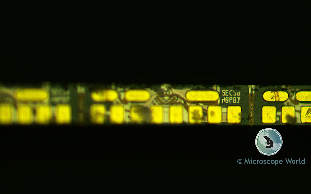
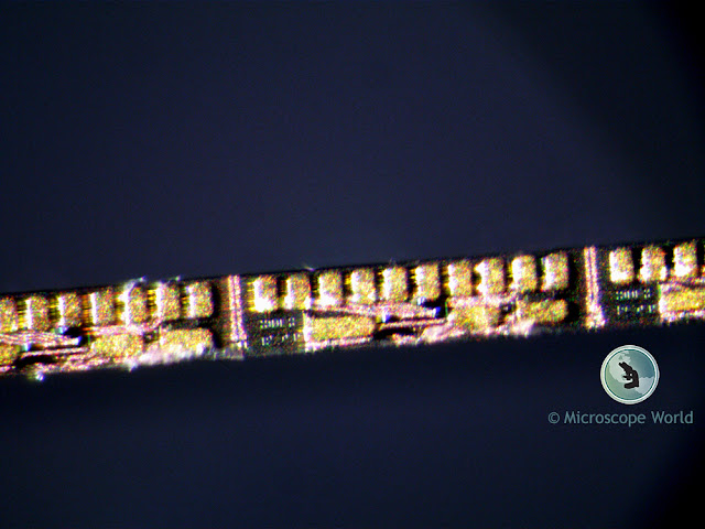
Below are two images of the same small printed part with text on it. The first image was captured using a stereo microscope with a lens that has NA of 0.10. This image was captured at 90x. Notice in the image below captured with the stereo microscope it is very hard to even read any of the printed text on the circuit. It should also be noted that it took nearly two hours to capture an image of this quality.

Image of a circuit captured under a stereo microscope at 90x magnification.
Stereo Microscope image captured at 90x, NA 0.10

The next image was captured using a metallurgical microscope with a lens that has NA of 0.30 and a magnification of 100x. This image took a few minutes to capture. The magnifications of the two captured images are similar however, notice how much easier it is to read the printed letters in the image that was captured with the metallurgical microscope. That ability is due to better resolution, which was obtained because of a higher numerical aperture of the lens used.

Metallurgical microscope image of a circuit captured at 100x magnification showing high quality resolution.
Metallurgical Microscope image captured at 100x, NA 0.30

Contact Microscope World with questions regarding NA, resolution, magnification or any other microscopy related questions.

Thursday, June 8, 2017

Corn Grain Under the Microscope

Corn is a large grain plant that was first domesticated by people in southern Mexico nearly 10,000 years ago. The ears of corn are actually fruit that produce the kernels (seeds) that people eat. Total production of corn worldwide surpasses that of wheat or rice. However, this is not all used for food. Corn is also used for producing ethanol, animal feed, corn starch and corn syrup.

The United states produced 361 million tons of corn in 2014, followed by China with 215 million tons and Brazil with 80 tons.

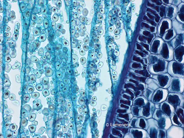
The images below of a cross section of kernel of corn were captured under the microscope using the Lumenera Infinity 3-3UR microscope camera.

Microscopy image of kernel of corn.
Grain of corn captured under the microscope with Infinity 3-3UR microscopy camera.

Microscope image of a kernel of corn.
Grain of corn captured under the microscope with Infinity 3-3UR microscopy camera.

Contact Microscope World for more information on Lumenera cameras or microscopes.

Friday, June 2, 2017

Middle School Microscopes

Microscope World offers several middle school microscopes that are perfect for kids exploring science!

MDS1 Middle School Microscope

Richter Optica MDS2 Middle School Microscope is a cordless and rechargeable microscope.
The Richter Optica MDS1 middle school microscope has several features that make it appealing to middle school classrooms and parents looking for a first microscope for their kids.
  • Cool, LED illumination (the bulb lasts a LONG time)
  • Carrying handle - perfect for younger students
  • Coarse & fine focusing, a must when viewing 400x
  • Cordless & Rechargeable - run the microscope corded or cordless
  • Retractable 40x lens helps teachers protect slides and the lens.

MDS2 Middle School Microscope

Richter Optica MDS2 Middle School Microscope has the ability to add a mechanical stage.
The Richter Optica MDS2 middle school microscope is a step up from the MDS1 in that it allows for the attachment of a mechanical stage. Here are a few features of the MDS2 middle school microscope. (Image shown includes optional mechanical stage).
  • Cool, LED illumination (the bulb lasts a LONG time) 
  • Coarse & fine focusing, a must when viewing 400x
  • Corded microscope
  •  Retractable 40x lens helps teachers protect slides and the lens.
  • Ability to add a mechanical stage, providing more precise control over X-Y movements of slides. 


MW2-HB4 Middle School Microscope

MW2-HB4 cordless middle school microscope from Microscope World.
The MW2-HB4 middle school microscope has a bit of a wider frame, providing added stability to the microscope.  A few features of this middle school microscope include the following:
  • Cool, LED illumination (the bulb lasts a LONG time) 
  • Coarse & fine focusing, a must when viewing 400x
  • Cordless & Rechargeable - run the microscope corded or cordless
  • Retractable 40x lens helps teachers protect slides and the lens.

Contact Microscope World with questions regarding middle school or student microscopes.