Thursday, April 27, 2017

Sickle Cell Anemia under the Microscope

Sickle cell anemia is an inherited form of anemia. It is a condition in which there are not enough healthy red blood cells to carry adequate oxygen throughout the body.

Normally, red blood cells are flexible and round, moving easily through blood vessels. When a person has sickle cell anemia, the red blood cells become rigid and sticky and are shaped like sickles or crescent moons. These irregularly shaped cells can get stuck in small blood vessels, which can slow or block blood flow and oxygen to parts of the body.

Microscopy image of sickle cell anemia at 40x magnification.
Sickle Cell Anemia under the microscope at 40x.

Symptoms of sickle cell anemia include fatigue, episodes of pain (when blood cells are being blocked), swelling of hands and feet, delayed growth, and vision problems.
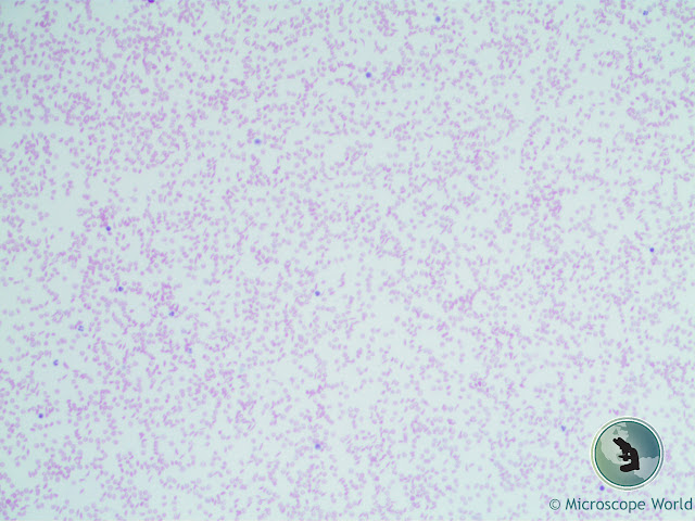
Microscope World image of sickle cell anemia under the microscope at 100x.
Sickle Cell Anemia under the microscope at 100x.

There is no cure for most people with sickle cell anemia. Bone marrow transplant or a stem cell transplant is the only potential treatment, but it is only used in children under age 16 because of the increased risk for older patients. Treatment for sickle cell anemia is usually aimed at avoiding crises, relieving symptoms and preventing complications.

These images are of sickle cell anemia cells that were captured under a lab microscope using a 5 megapixel microscopy camera.
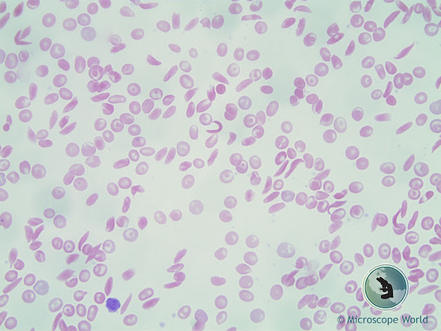
Microscope World image of sickle cell anemia captured at 400x.
Sickle Cell Anemia under the microscope at 400x.

Learn more about sickle cell anemia here.

Contact Microscope World with questions regarding microscopes and microscopy cameras.

Tuesday, April 25, 2017

High School Binocular Microscope

The Richter Optica HS-3B is a versatile high school compound microscope. A few features make this microscope one of the most popular microscopes across high schools in the United States.
  • Binocular eyetubes with WF10x eyepieces.
  • 40x, 100x, 400x, 100x magnification.
  • Retractable 40x and 100x (oil) objective lenses avoid damage if moved into contact with the stage.
  • Frame provides easy carrying handle, with cord storage behind microscope.
  • Operates corded or cordless.
  • Rechargeable batteries.
  • LED light is cool and provides long-lasting bulb.
  • Mechanical stage is attached and locked on.
  • Coarse and fine focusing allows for a crisp and clear image.
  • 100~240V allows for use in all countries.
  • Microscope is also available in pared down 400x magnification model, HS-3B-3


HS-3B Student Microscope
Richter Optica HS-3B Microscope

You can view all the Richter Optica microscopes and accessories here. Contact Microscope World for a quote.

Tuesday, April 18, 2017

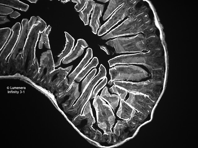
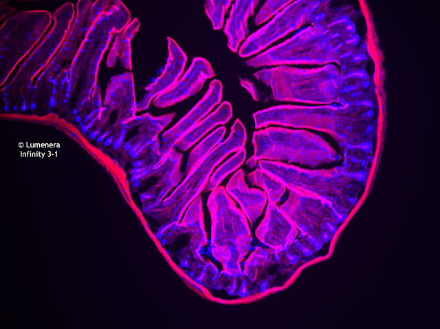
Mouse Intestine under the Microscope

The images below of a mouse intestine were captured using a Lumenera Infinity 3-1 microscope camera. This is a cooled, CCD camera that is great for low-light situations or where a high dynamic range is required. This 1.4 megapixel camera is ideal for the following applications:
  • Fluorescent Microscopy
  • Green Fluorescent Protein Applications
  • Fluorescent In Situ Hybridization
  • DNA Analysis
  • Live Cell Imaging
  • Brightfield, Darkfield, DIC/Phase Contrast
  • Near IR Applications
  • Histology, Pathology and Cytology
  • Forensic Analysis
  • Semiconductor Inspection
  • Metallurgical Microscopy 
Mouse intestine under the microscope captured with Lumenera Infinity 3-1 CCD camera.
Mouse intestine captured with the Monochrome Infinity 3-1 microscopy camera.

Mouse intestine captured with the Color Infinity 3-1 microscopy camera.

Infinity 3-1 microscopy camera image of mouse intestine under the microscope using fluorescence.
Mouse intestine captured with the Color Infinity 3-1 microscopy camera.

Contact Microscope World for more information on microscopy cameras.

Friday, April 14, 2017

Hair Under the Microscope

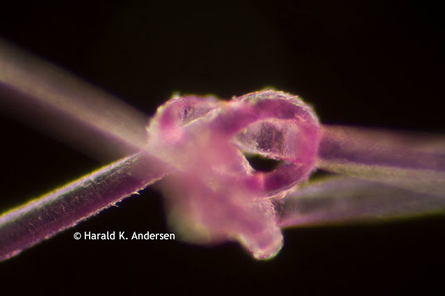
Harald K. Andersen of Steinberg, Norway captured these images of hair under the microscope. The first image is of a single strand of hair captured through the microscope at 20x magnification. The hair was dyed for the project.

Hair captured under the microscope.
(C) Harald K. Andersen. Single strand of hair under the microscope.

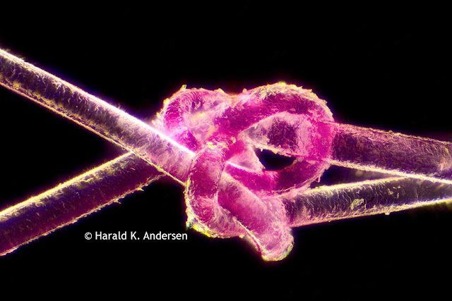
In order to capture the next image Andersen stacked together 480 images using a software called Zerene Stacker.

Hair captured under the microscope.
(C) Harald K. Andersen. 480 stacked images of a single strand of hair under the microscope.

Andersen mentioned that the hardest part of the project was tying the knot in the hair! A big thanks for sharing the images with Microscope World!

Friday, April 7, 2017

Tonsils under the Microscope

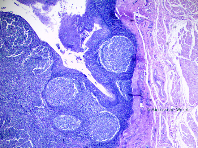
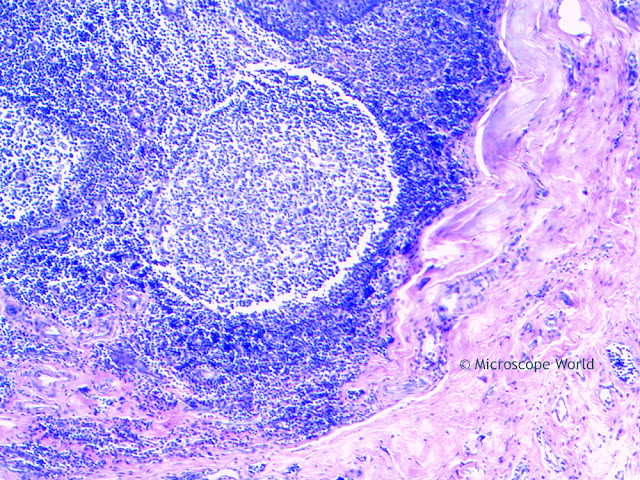
The palatine tonsils are two lymphatic masses, located at the back of the throat on each side of the oral pharynx. The tonsils play a role in protecting the body against respiratory and gastrointestinal infections.

Each tonsil consists of a network of crypts (pits) that store cells used to fight infection. The tonsils contain B cells, a type of white blood cell that fights infections. They also produce antibodies against polio, streptococcal pneumonia, influenza, and numerous other infections. Antibodies are proteins that help the body identify and attack harmful invaders.

The tonsils also contain several types of T cells, which are white blood cells that destroy cells infected with viruses and help the body build immunity to infectious organisms.

The images below are cross sections of Palatine Tonsils that were captured using a biological lab microscope and a microscopy camera.

Microscopy image of tonsil under the microscope at 40x captured by Microscope World.
Palatine tonsil under the compound microscope at 40x.

Microscope World image of palatine tonsil cross section under the microscope at 100x.
Palatine tonsil under the compound microscope at 100x.

Microscope World image of palatine tonsil cs captured at 400x.
Palatine tonsil under the compound microscope at 400x.

Contact Microscope World with any questions about microscopy or capturing digital images under the microscope.

Tuesday, April 4, 2017

Worms found in Cows under the Microscope

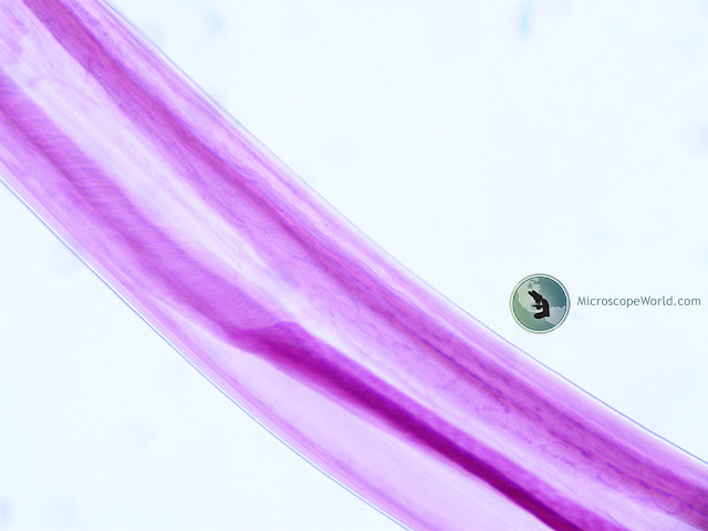
Bunostomum phlebotomum is a species of worms (nematodes) that are found in the small intestine of calves. The worms are stout and measure 1-3cm in length. Young animals are most commonly affected and only several hundred worms can cause death. Adults often carry worms but do not show any symptoms.

Diarrhea is the primary symptom in the cows along with signs of anorexia, lethargy and weight loss. Hypoproteinaemia, anaemia and dehydration often occur. Skin involvement due to larvae penetration appears as lesions especially on the feet and limbs. Analysis of fecal samples under a microscope enables eggs to be found, or adult worms can be visualized in the small intestine when an autopsy is performed.

Veterinarians and ranchers can use Anthelmintics (antiparasitic drugs) for effective treatment of the infection. Preventative antiparasitic treatment may help prevent the disease as well as pasture management to avoid egg and larvae accumulation.

The image below is Bunostomum phlebotomum that was taken from a calf in Texas. Images were captured using a 3 megapixel microscopy camera on the RB20 Lab Microscope.

Microscope World image of bunostomum phlebotomum under the microscope at 100x.
Bunostomum Phlebotomum (worm) taken from a cow's intestine, examined under the RB20 Microscope at 100x.

Microscope World image of Bunostomum phlebotomum under the microscope at 400x.
Bunostomum Phlebotomum (worm) taken from a cow's intestine, examined under the RB20 Microscope at 400x.