Tuesday, March 28, 2017

Museum Display Microscope

The museum demonstration high definition display microscope was designed for use in museum and art exhibits. The stereo microscope provides dual magnification of 10x and 30x, ideal for viewing archeological samples, botany samples, fossils and insects. The microscope is equipped with a high definition HD camera and a 12" HD monitor.

The high definition monitor can face any direction, providing some options for exhibit setup. If the monitor is facing away from the eyepieces the microscope can be set up allowing visitors to look through the eyepieces on one side of the exhibit, while on the other side guests can gather around the monitor and view the samples.

Alternatively, the museum microscope could be set up in an area where visitors don't have the ability to touch the microscope, but are still able to view the samples underneath the microscope.

The display microscope has top and bottom LED illumination. This light is a cool light and will not heat up or harm any living specimens. Each light has its own rheostat intensity control.
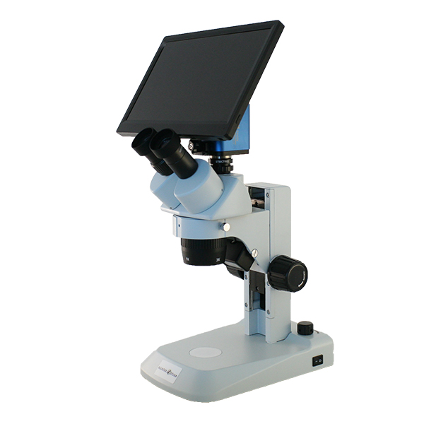
Museum display microscope with HD digital camera.
Museum Display HD Microscope

Wednesday, March 22, 2017

Intestine under the Microscope

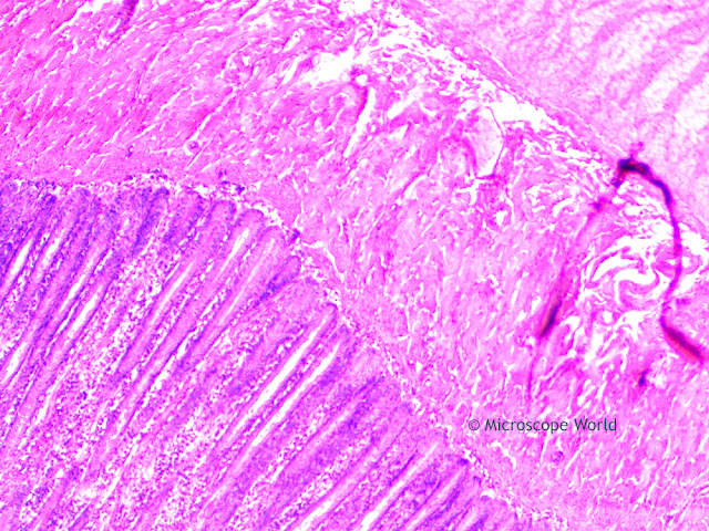
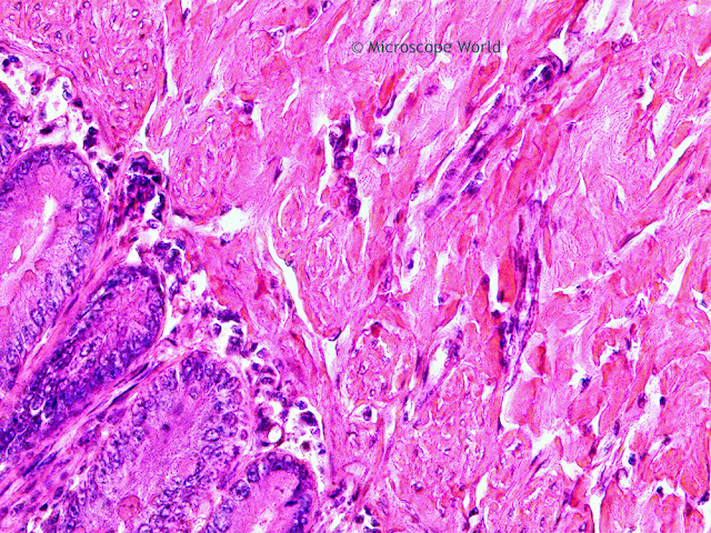
The Jejunum is the middle portion of the small intestine, connecting the duodenum and the ileum. It is partially responsible for absorbing nutrients into the bloodstream. It is lined with finger-like projections, that are called villi, that move nutrients, vitamins and minerals from the intestine into the bloodstream where they can be used by the entire body.

The images below were captured with the RB30 lab microscope using a high definition HD microscopy camera.

Microscope World image of the jejunum captured under the microscope at 40x.
Jejunum under the microscope at 40x.

Microscope World image of the small intestine captured at 100x under the microscope.
Jejunum under the microscope at 100x.

Microscope World image of the Jejunum (small intestine) under the microscope at 400x.
Jejunum under the microscope at 400x.

Contact Microscope World with questions about digital microscopy.

Wednesday, March 15, 2017

Images captured with HS2 High School Microscope

Richter Optica HS-3B Microscope

The Richter Optica HS3 is available as a monocular microscope (HS-3M) or as a binocular microscope (HS-3B). The microscope comes standard with 4x, 10x, and 40x Achromat objective lenses and an optional 100x lens is available on some models. This microscope is used most commonly in high schools.

The images below were all captured using the 40x achromat objective lens that comes on the HS-3 microscopes. A 5 megapixel microscope camera was used to capture the images.


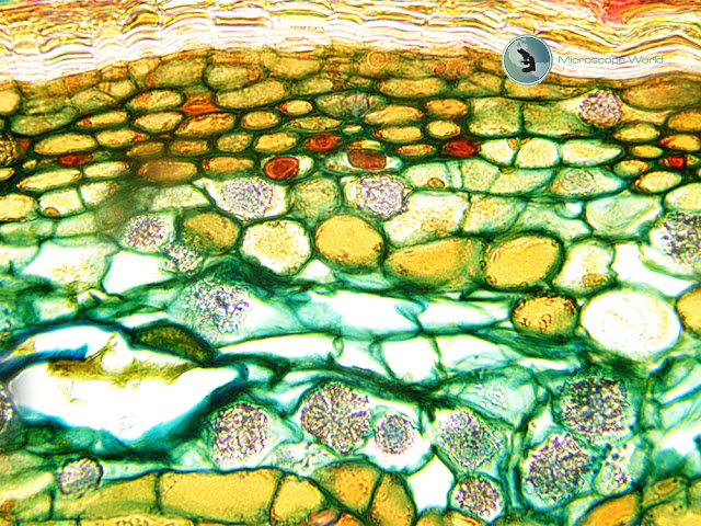
Microscope World image of Tilia under the microscope at 400x.
2-Year Tilia captured at 400x with the HS3 High School Microscope

Microscope World image of monocot and dicot at 400x.
Monocot & Dicot captured at 400x with the HS3 High School Microscope

Microscope World image of plant cells captured at 400x magnification under a high school microscope.
Plant Cells captured at 400x with the HS3 High School Microcope

Contact Microscope World for more information on high school microscope options.

Thursday, March 9, 2017

Microscope Service

Microscope World provides microscope service and microscope maintenance in Southern California. With over 30 years of technical experience, your microscope will be in good hands whether you need routine maintenance or need a specific problem fixed. You rely on your microscope to perform on a daily basis and if a problem should arise Microscope World understands the number one priority is to get your instrument back up and functioning properly in a timely manner.

  • Parts available for most microscope brands.
  • Volume discounting.
  • Over 30 years of technical experience.
  • Access to parts that are no longer supported by manufacturers.
  • Certified instrument calibrations.
  • Factory trained technicians.
Learn more about Microscope World's microscope servicing here.

Tuesday, March 7, 2017

Textile Exam Microscope for Methods AATCC 20 and 20A

The Association of Textile, Apparel, and Materials Professionals has standards that are required for different test methods. There are several test methods that require the use of a microscope for examining textiles.
  • Test Method AATCC 20 is a qualitative fiber analysis test.
  • Test Method AATCC 20A is a quantitative fiber analysis test.
Microscope World has a specific microscope that meets test method AATCC 20 and AATCC 20A standards for textile examination.

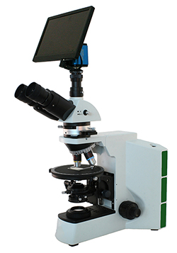
Textile Exam Micrsocope for Method AATCC 20 and AATCC 20A
Textile Exam Microscope
The AATCC 20 and AATCC 20A textile examination microscope includes the following:
  • Polarizer and Analyzer
  • Gypsum Full λ Wave Plate
  • Mica 1/4 λ Wave Plate
  • Infinity Corrected Optics
  • Stress Free Plan Achromat 4x, 10x, 40x, 60x Objective Lenses
  • High Definition Camera with Image Capture to SD Card
  • 12" HD Monitor that Connects to Camera
  • Mechanical Stage
Learn more about the AATCC 20 and AATCC 20A textile exam microscope here.