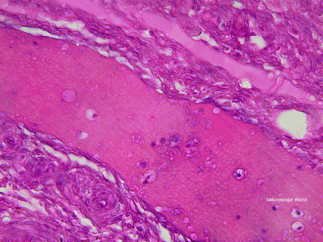
The ovary is one of two reproductive glands in women. The ovaries are located in the pelvis, one on each side of the uterus. Each ovary is about the size and shape of an almond. The ovaries produce eggs (ova) and female hormones. During each monthly cycle, an egg is released from one ovary. The egg travels from the ovary through a fallopian tube to the uterus. The ovaries are the main source of female hormones, which control the development of female body characteristics, such as the breasts, body shape and body hair. They also regulate the menstrual cycle and pregnancy.
The images below of an ovary were captured with the
RB30 lab microscope using the
DCM5 5 megapixel microscope camera and software. The first three images were captured using Plan Achromat objective lenses, and the final image was captured using a
Plan Semi Apochromat Fluor 40x objective lens.
Contact Microscope World for further information on microscopy solutions.


