Monday, April 25, 2016

Apple under the Microscope

The apple is the fruit from the apple tree, a deciduous tree in the rose family. The apple tree originated in the Middle East about 4,000 years ago, and is one of the oldest known fruits.

As with the old adage, "An Apple a Day Keeps the Doctor Away" the apple is a powerhouse of nutrients with a great source of fiber, vitamin C, heart-healthy potassium, B-complex vitamins, phytonutrients, antioxidants, and many more health and medicinal benefits.

The vitamins present in apples are the key in maintaining red blood cells and keeping the nervous system in good health. The nutrients in apples are unduly present in the skin of the apple, which is the most valuable part of the fruit with respect to its nutrient substance.

The images below are of a cross section of an apple on a prepared slide under the UX-1D plan achromat digital microscope.

Microscopy image of an apple under the microscope at 40x.
Apple under the UX-1D digital microscope at 40x

Microscpy image of an apple at 100x.
Apple under the UX-1D digital microscope at 100x

Micorsocpy image of an apple at 400x by Microscope World.
Apple under the UX-1D digital microscope at 400x

For more information on digital microscopes contact Microscope World.

Thursday, April 21, 2016

8 Tips to Capture Great Microscopy Images Before the Fluorescence Fades

Fluorescence microscope image.Fluorescence is a great technique for microscopy. It provides amazing contrast and allows us to selectively view the objects that interest us without being obscured by surrounding structures. The one big drawback it has over brightfield microscopy techniques is bleaching. Even the brightest fluorescently labeled samples will eventually fade. However, there are several things you can do to prolong the effective fluorescence and capture some great images before they fade into oblivion.

  1. Use DIC or Phase Contrast to find your sample.
    After you place your sample on the stage, don't switch to fluorescence immediately. Use brightfield illumination such as phase contrast or DIC to find a suitable area to image. Once you find the right spot you can take a quick peek with fluorescence to make sure the fluorescent label is visible.
  2. Power down.
    Unless you are using very dim fluorophores, you most likely will not need the full power of your UV light source. Most LED based light sources allow you to adjust the light intensity. Start with the lowest intensity and gradually increase it until you can just see the structure you want to image (it doesn't have to be that bright - remember that your microscopy camera will pick up a lot more light than your eyes do). On confocal or other laser-based systems a laser power of just a few percent is often sufficient for a well-labeled sample. If you are using a mercury or xenon lamp, consider using an attenuator to limit the amount of light reaching the sample.
  3. Tune your camera settings.
    Dedicated microscope cameras have been optimized for fluorescence imaging and have many features that will allow you to collect data as quickly as possible before your sample fades. Avoid color cameras as the Bayer mask in front of the chip absorbs a significant amount of light. Check your camera's operating manual for settings such as gain, binning modes, sub-frame readout, and dynamic range. All of these can be adjusted to increase image capture speed but there is usually some cost in terms of noise or resolution. Some camera control interfaces allow you to use high-speed features in a preview mode (for example, while you’re focusing the sample) and then automatically switch to a high quality mode to capture the image. It is a good idea to test camera settings on a positive control or spare sample before imaging your critical experiment.
  4. Use objectives with high numerical aperture values.
    The higher the numerical aperture of the objective, the more light will be collected from the sample which, in turn, allows you to reduce the exposure time. Find the objective with the highest numerical aperture that will still give you the field of view that you need. If available, use oil immersion objectives as they typically have a higher NA than comparable dry lenses.
  5. Avoid z-stacks or autofocus.
    If you have a motorized Z-drive on your system, you may be tempted to use some of the enhanced functions that this feature offers such as autofocus and Z-stack imaging. While these may be necessary if you are doing timelapse imaging or you have thick specimens, keep in mind that you are prolonging the exposure of your sample to fluorescent light when you use these features.
  6. Use bright, stable fluorophores.
    There is considerable variability in the brightness and stability of many of the fluorophores typically used in fluorescence microscopy. Before you grab the first fluorescent dye you can find in your freezer, take a moment to look up the quantum yield (brightness) and photostability of the dye. These may not always be easy to find but picking the right fluorophores can save you a lot of trouble later on.
  7. Use an anti-fading agent.
    Using an anti-fading agent in your mounting medium can dramatically decrease the speed of bleaching in your sample. There are some home brew recipes available but commercial products such as ProLong and VECTASHIELD are not too expensive and work extremely well.
  8. Use dedicated, high-efficiency filter sets.
    Many fluorescence microscopes come with a standard filter set for imaging red, green, and blue fluorescence. These work well enough for routine imaging with a variety of common fluorophores such as DAPI, FITC, TRITC, Texas Red and even GFP. That versatility comes at a cost, though, as these filter sets usually don’t have the highest transmissions and the broad band pass values can cause bleed-through when imaging dyes that are spectrally close. If you know you will be doing a fair amount of imaging with a particular fluorophore, it is probably worthwhile investing in a dedicated, high-efficiency, filter cube for that dye.

If you have questions regarding fluorescence microscopy please contact Microscope World.

Wednesday, April 13, 2016

Wildberry and a Guest Insect

Microscope World was recently capturing some images of some wild berries using the S6D-BL digital stereo zoom microscope. While capturing the images we noticed a guest on one of the berries. Look at the berry in the top right of the screen - this insect must be loving his home/food!


Friday, April 8, 2016

Ovary under the Microscope

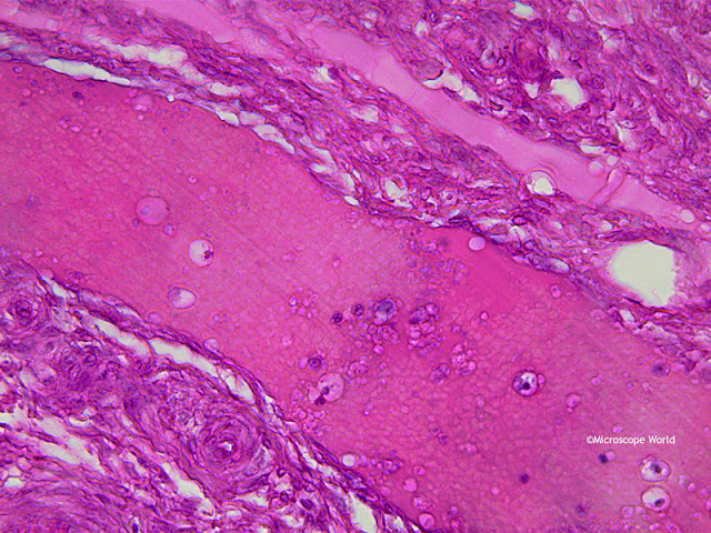
The ovary is one of two reproductive glands in women. The ovaries are located in the pelvis, one on each side of the uterus. Each ovary is about the size and shape of an almond. The ovaries produce eggs (ova) and female hormones. During each monthly cycle, an egg is released from one ovary. The egg travels from the ovary through a fallopian tube to the uterus. The ovaries are the main source of female hormones, which control the development of female body characteristics, such as the breasts, body shape and body hair. They also regulate the menstrual cycle and pregnancy.

The images below of an ovary were captured with the RB30 lab microscope using the DCM3 3.2 megapixel microscope camera and software. The first three images were captured using Plan Achromat objective lenses, and the final image was captured using a Plan Semi Apochromat Fluor 40x objective lens.

Microscopy image of an ovary at 40x magnification.
Ovary captured under a lab microscope at 40x magnification.

Image of an Ovary under a lab microscope at 100x magnification.
Ovary captured under a lab microscope at 100x magnification.

Microscope image of an ovary captured by Microscope World.
Ovary captured under a lab microscope at 400x magnification.

Microscopy image of ovary under the microscope at 400x magnification, captured by Microscope World.
Ovary captured under a lab microscope at 400x magnification using a Plan Semi Apochromat Objective.


Contact Microscope World for further information on microscopy solutions.

Monday, April 4, 2016

Huvitz HRM-300 3D Profiler Microscope System

The world of microscopy is a fascinating place and using digital technologies such as 3D profiling, are allowing us to analyze samples in ways that were extremely difficult and sometimes not possible in the past.

I decided to get familiar with the Huvitz HRM300 series metallurgical microscope and its Panasis 3D profiling system software to see what could be done with this technology and how user friendly it is. I used the LUSIS HC-20CU microscopy camera that is included with this package to capture images and video of a circuit board.

Microscope 3D Image profiling system HRM300 from Microscope World
Huvitz HRM300 3D Profiler System with Camera, Control Box and 3D Imaging Software

Microscope mechanical stage with circuit board.
Circuit Board on 100mm x 100mm Travel Mechanical Stage

Huvitz 3D profiler Panasis software and control pad.
Panasis Interface on left monitor, User's Manual on right and Control Pad
HASP Key & USB to RS232 Adapter

There were some basic configurations and a calibration that I needed to perform, but afterward, and with a little help from the manual's step-by-step instructions, I was ready to take an Extended Depth of Focus photo of the circuit board.


Panasis extended depth of focus software microscopy circuit board image.
Panasis Extended Depth of Focus Image of the Circuit Board

Using the mechanical stage and viewing the live image on the monitor was simple enough and soon I found the area that I wanted to view. Under the 3D Profiler tab I set the top and bottom image reference points for the image and on the Extended Depth of Focus (EDF) tab I selected Auto and then entered the height intervals to be taken between image stacks. I then clicked Start and sat back while the profiler did its work.

The Huvitz HRM-300 microscope's stage began to move between the top and bottom limits of the z-axis with a quiet motorized sound. A few seconds later a very detailed and crisp image of the circuit board appeared on the screen. I was impressed by the clarity of the image, but now it was time to see it in 3D. So I clicked on the 3D icon that appeared in the top right corner of the image and . . . 

Microscopy 3D image of circuit board captured under HRM300 Huvitz metallurgical microscope with 3D profiling.
3D Image of Circuit Board under the Microscope!
The Panasis software allows for all types of measurements to be conducted so that the data and statistics needed are a few simple selections away.

Microscope 3D image of circuit board with image texture blending to see sample's surface at designated ratios.
Image Texture Blending to help see sample's surface at designated ratios.

Making profile measurements with the Huvitz HRM-300 metallurgical microscope with 3D profiling.
Making Profile Measurements

Huvitz HRM-300 3D Image profiler software showing volume measurements of a circuit board.
Volume / Area Measurements

Surface analysis microscope with 3D imaging software.
Step Measurements & Lighting Enabled with Histogram for Convenient Surface Analysis.

The overall user interface was pretty straight forward and the manual and other support options for the Huvitz HRM-300 series microscope, Panasis 3D profiling imaging software, and the camera made for a productive and user-friendly experience.

There are a lot of other cool features that come along with this microscope 3D profiling system including the following:
  • The ability to obtain wide panorama imaging of your sample, which greatly extends the microscope's field of view.
  • Automatic optimized contestant lighting when observation magnifications are changed.
  • Environment friendly functions that are good for both your microscope, the sample and the environment. The ECO setting goes into auto-power saving mode when you walk away from the system. Bulbs last longer and energy is saved. You can adjust the sleep mode time through the software.
  • Additional options are available including a variety of objectives, a motorized nosepiece, motorized stage and reflected and transmitted illumination.
 Do you have experience using the Huvitz HRM-300 3D Profiler Microscope? How has it worked for you and what are your thoughts on the 3D digital microscopy technology? Microscope World would love to hear from you regarding with questions or comments on this microscope system. Contact Microscope World and ask specifically for microscopy specialist Sean Page. Stay tuned for more of Sean's adventures in microscopy at Microscope World.