Tuesday, November 24, 2015

Dissecting Microscopes

What exactly is a dissecting microscope? There are several different types of dissecting microscopes and we will cover each of them below. A dissection microscope is also referred to as a stereo microscope or a low power microscope.

A few features common among all dissecting microscopes:
  • Dissecting microscopes have working room on the stage. There is room to place larger objects such as rocks or nuts and bolts. In order words, the microscopy sample does not need to be on a microscope slide.
  • Dissecting microscopes use light from above the stage, making it possible to view opaque objects. Some dissecting microscopes also have light beneath the stage, but not always.
  • Dissecting microscopes provide low magnification (usually 10x - 40x is a common range) and they are used to view detail in objects you can already see with the naked eye. Blood cells are not viewed with a dissecting microscope, but rather with a compound microscope.

Single Power Dissecting Microscopes

Single magnification stereo microscope MW1-L2.
Single Magnification Microscope

Single power dissecting microscopes offer one single magnification. For example, the microscope might provide 20x magnification. These single power instruments are common in museums or schools where only one magnification is required to view samples. With a single power magnification dissection microscope it is possible to have two magnifications when using a different pair of eyepieces. For example, with a 10x and 15x pair of eyepieces (when the built-in objective is 2x) total magnification would be 20x and 30x. This is an inexpensive way to achieve multiple magnifications from a lower cost microscope.

Dual Power Dissecting Microscopes

Stereo microscope with dual magnification.
S2 Dual Magnification Microscope

Dual power dissecting microscopes provide two set magnifications. By turning a turret on the bottom of the body of the microscope (the black portion on the image at right), it is possible to flip back and forth between the two magnifications. The objective lenses are built into the microscope (for example, 1x and 3x) and the eyepieces can be changed. On a microscope that includes 10x eyepieces with a built-in 1x and 3x objective, the total magnification is 10x and 30x. Magnification could be increased by purchasing a pair of 15x eyepieces, which would provide 15x and 45x magnification. These microscopes are sometimes used by coin collectors, in schools, and in industrial quality control settings where an inexpensive microscope is required.

Zoom Dissecting Microscopes 

Stereo zoom dissecting microscope.
Stereo Zoom Microscope
Stereo zoom microscopes provide a magnification range and all magnifications within this range can be viewed. The S6 stereo microscope shown at left provides magnification of 6.7x - 45x, which means you can see 10x, 15x, 18x, etc. all the way up to 45x. These are the most commonly used dissecting microscopes, since they provide more options for viewing the exact part of the sample at the desired magnification. Typically when increasing magnification on a stereo zoom dissection microscope, rather than changing eyepieces, an auxiliary lens is added to the bottom of the stereo microscope body. If you were using the microscope shown at left and wanted a bit more magnification, you might add the 1.5x auxiliary lens, which would change the magnification range to 10x - 67.5x. Stereo zoom microscopes are typically used in universities, in industrial settings for quality control and to view small parts, and in manufacturing.

Friday, November 20, 2015

Insects Under the Microscope

Microscope World recently wanted to compare two different microscopy cameras for quality and differences in color. The two microscope cameras included the DCM5 documentation camera (5 megapixels) and the HDCAM4 high definition camera (2 megapixels). The onscreen resolution and image quality from the HD camera is amazing, so there was a question as to how the captured image would match up to the onscreen quality.

 To give you an idea of size, this is the insect that was placed under the microscope.

Insect used under the microscope.

The images below were captured using the S6.7 Trinocular Stereo Zoom Microscope.

Insect captured with S6T-LED stereo microscope and DCM5 Microscope Camera at 6.7x.

Insect captured with S6T-LED stereo microscope and HDCAM4 Microscope Camera at 6.7x.

Insect captured with S6T-LED stereo microscope and DCM5 Microscope Camera at 20x.

Insect captured with S6T-LED stereo microscope and HDCAM4 Microscope Camera at 20x.

The color in the DCM5 microscope camera are a bit brighter than those in the HD microscope camera, but overall the image quality was similar. If you have questions regarding microscope cameras please contact Microscope World.

Wednesday, November 18, 2015

Top 10 Gift Ideas for Science Enthusiasts

Whether you are shopping for a graduation gift, birthday, or a holiday, science enthusiasts can be tricky to shop for. The list below includes some ideas for both younger and grown-up scientists. Shopping for science geeks just got a bit easier!
Freudian Slippers

Tuesday, November 10, 2015

Kids Science Project: Fungus Growth

Bacteria, fungi and other microscopic organisms that live in soil, air and water are responsible for turning once-living plants and animals into nutrients that can be used again. Decomposition may seem gross when you find bread molding in the fridge, but it is actually a fundamental process on which all life depends! Fungi are able to produce special enzymes that allow them to break down dead plants and animals and use them as food.

Growth of bacteria and fungi can occur at an amazingly fast rate. In four hours one bacterium can grow to a colony of over 5,000. In just one teaspoon of soil there are more bacteria and fungi than the number of people on earth!

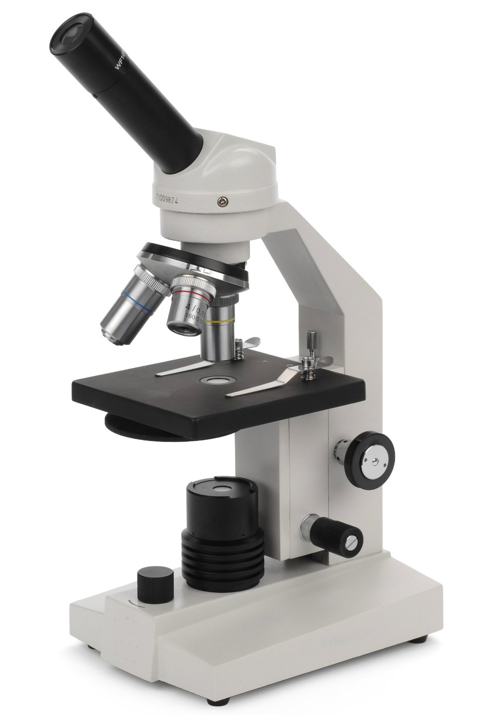
This kids science project involves growing fungus on bread, observing it as it grows, and then viewing the bacteria under a dissecting microscope (low magnification microscope).

Items required:
  • 3 Petri Dishes
  • 3 Ziplock bags
  • 3 Slices of bread 
  • Two different types of soil (from different areas or gardens)
  • Stereo Microscope
Place a slice of bread on a flat surface and press the petri dish into the bread, removing the excess. Do this for each petri dish. Mix two bowls of soil with water. Place about ten drops of soil water on two of the bread samples (the third sample does not get any soil - it is your control group). Place each petri dish inside a ziplock bag and seal the bag. Make sure you label them! Place the sealed bag petri dishes into brown paper bags and put them in a warm, dark environment to promote bacteria and fungi growth.

Each day remove the petri dish from the bag and observe any mold growth under the microscope. Write down your findings and draw or capture images. Are the colonies of bacteria or fungi different sizes and colors? Is one bread petri dish growing faster than another?

Microsocpy image of Funghi spores.
Funghi spores under the microscope.

Definitions for students writing science reports on this experiment:

Bacteria - a widely distributed group of typically one-celled microorganisms, some of which produce disease. Many are active in processes of fermentation, which is the conversion of dead organic matter into soluble food for plants.

Decomposition - organic decay.

Decomposers - organisms which break materials down into parts and cause them to rot.

Enzymes - any of a number of proteins or conjugated proteins produced by living organisms and functioning as biochemical catalysts.

Fungus - any of a number of plants lacking in chlorophyll, including yeasts, molds and mushrooms.

Wednesday, November 4, 2015

48 New Land Snail Species Discovered

Researchers recently added to their knowledge of land snails of Sabah (Malaysia, Borneo) including the discovery of 48 new species of land snails. Jaap J. Vermeulen, Thor-Seng Liew and Menno Schilthuizen reported on their findings in a full abstract here. Land snails were collected in large quantities from a variety of areas including quarries, river beds and on the side of the road. The land snails were then examined and viewed under a stereo microscope.


The drawings of the land snails below were created using a stereo microscope along with a camera lucida device to aid in drawing. When naming the new snails, in the enumerations of localities, words were used from the Malay language: batu (= rock), bukit (= hill), gua (= cave), gunung (= mountain), pulau (= island), and sungai (= river).

Stereo microscope image sketch of land snails found in Malaysia
Sketch courtesy: Additions to Knowledge of Land Snails Research Article

The land snails labeled 1 are Acmella cyrtoglyphe sp. 1A & 1B were found in the Sepulut Valley in the Interior Province of Sabah, Malaysia. 1C was found in Kinabatangan Valley in the Sandakan Provice.
The land snails labeled 2 are Acmella umbilicata sp. and were found in the Pinangah Valley in the Interior Provice of Sabah, Malaysia.
  • Land snail 1A is 1.4mm high and when viewing this land snail with a stereo microscope at 50x magnification, the land snail would fill about 30% of the field of view
  • Land snail 2A is 1.3mm high and when viewing this land snail with a stereo microscope at 50x magnification, the land snail would fill about 28% of the field of view.

 
Land snails from Malaysia under a stereo microscope.
Sketch courtesy: Additions to Knowledge of Land Snails Research Article
The land snails labeled 9 are Ditropopsis davisoni sp. and were found in the Matang River South of Long Pasia in the Upper Padas in Sabah, Malaysia.
The land snails labeled 10 are Ditropopsis trachychilus sp. and were found on Kota Kinabalu-Tambunan road in Crocker Range N.P. in Sabah, Malaysia.
The land snails labeled 11 are Ditropopsis koperbergi and were found in the Danum Valley Conservation Area, Sabah, Malaysia.
  • Land snail 10A is 2.3mm high and when viewing this land snail with a stereo microscope at 50x magnification, the land snail would fill about 50% of the field of view

Small land snail under the microscope!
Sketch courtesy: Additions to Knowledge of Land Snails Research Article


The land snails labeled 3 are Acmella polita and were found in the north end of the limestone ridge on the East bank of the Tabin River in the Segama Valley, Sandakan Province, Sabah, Malaysia.
The land snails labeled 4 are Acmella ovoidea sp. and were found in the Pinangah Valley in the Interior Province of Sabah, Malaysia.
The land snails labeled 5 are some of the smallest land snails discovered: Acmella nana sp. and were found in the Niah Caves on the west side of the quarry in Sarawak, Malaysia.
  • Land snail 5A is 0.7mm high and when viewing this land snail with a stereo microscope at 50x magnification, the land snail would only fill about 15% of the field of view

Monday, November 2, 2015

Mixing Microscope Objective Lenses

Is it possible to mix and match objectives lenses from one brand of microscope to another? It depends on your type of microscope, but sometimes yes, it is possible. The objective lenses you use on your microscope need to be the same tube length as the microscope. For example, if your microscope has a 160mm fixed tube length you will need to use 160mm tube length objective lenses. Many older or inexpensive microscopes, when measuring from the back end of the microscope to the primary focal plane are limited to 160mm, therefore they have a 160mm fixed tube length. More advanced microscopes use a series of lenses and prisms to allow for an "infinite" distance between the back end of the microscope to the primary focal plane. These microscopes have an "Infinity Corrected" optical system and will use Infinity Corrected objective lenses.

Microscope Objective Lenses
A few things to keep in mind if you do choose to mix and match microscope objective lenses from one microscope brand to another.
  • Digital microscopy images may not match up when mixing objective lenses from one brand to another, even if you make sure the microscope tube length and the objective are comparable. You may be able to simply refocus the microscopy image for the camera.
  • If you are using mixed objective lenses on the microscope turret, they most likely will not be parfocalled (each objective in focus when switching from one magnification to another). You can still use the objectives, just be prepared to allow a bit more time for re-focusing when changing magnifications.
  • Before you purchase another objective lens, make sure the lens uses the same thread size or be prepared to use a threaded adapter.
  • If you are using specialty microscopy techniques such as phase contrast or darkfield microscopy mixing objective lenses might not work well.