Thursday, September 25, 2014

Ranunculus under the Microscope

Ranunculus are a colorful genus of flowers that include buttercups, spearworts, water crowfoots and celandine. Typically Ranunculus have five petals, but occasionally more.
 
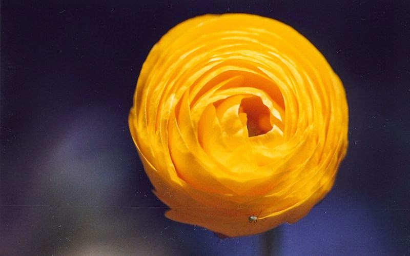
Yellow Flower
Ranunculus image courtesy of Joydeep Sen Sarma


The name Ranunculus is Latin for "little frog" and might refer to the many species of flowers found near water, similar to frogs.

The images below of Ranunculus were captured under the U2 biological microscope using  a 5 megapixel microscope camera.

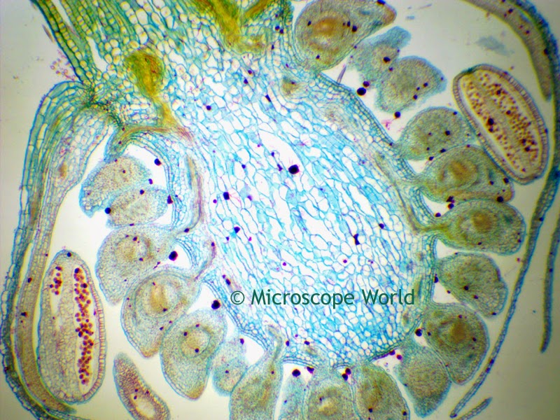
Ranunculus under the microscope at 40x
Ranunculus under the microscope at 40x magnification.

The Ranunculus prepared microscope slide is part of the Fruit and Flower microscope prepared slide kit available from Microscope World.

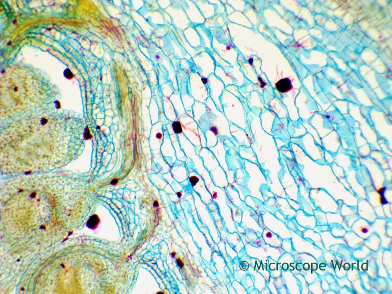
Ranculus under the microscope at 100x magnification.
Ranunculus under the microscope at 100x magnification.
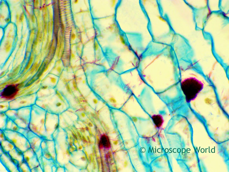
Ranculus under the microscope at 400x magnification.
Ranunculus under the microscope at 400x magnification.

Monday, September 22, 2014

Minerals Under the Microscope

Minerals are best viewed under a stereo zoom microscope. A customer of Microscope World's, Mr. Arnie Lambert captured these mineral images using a stereo microscope and a Nikon SLR camera. The Nikon SLR camera was connected to the microscope through the eyetube on his microscope using the Nikon SLR camera microscope adapter.
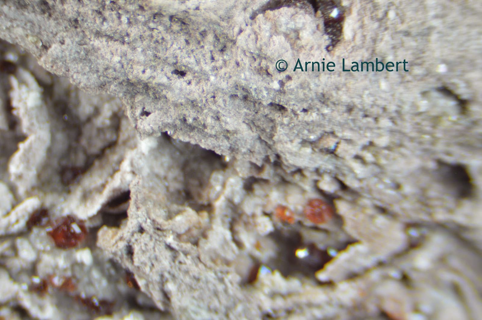
Garnets in Rhyolite under the microscope, from New Mexico
Garnets in Rhyolite from New Mexico.
Garnets are a group of silicate minerals that have been used for many years as gemstones and abrasives. Garnet can be a variety of colors including red, orange, yellow, green, purple, brown, blue, pink, colorless and even black. The rarest color is blue. Rhyolite is an igneous, volcanic rock.
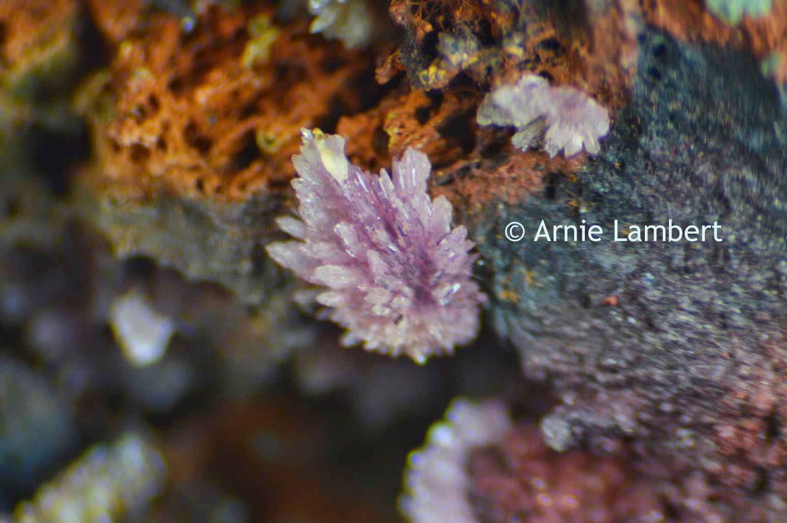
stengite under microscope from Indian Mountain
Strengite from Indian Mountain in Cherokee Co., Alabama
Stengite is a relatively rare iron phosphate mineral that is lavender, pink, colorless, or red. This mineral was named after Johann August Streng (1830-1897), a professor of Mineralogy at the University of Giessen, Germany.
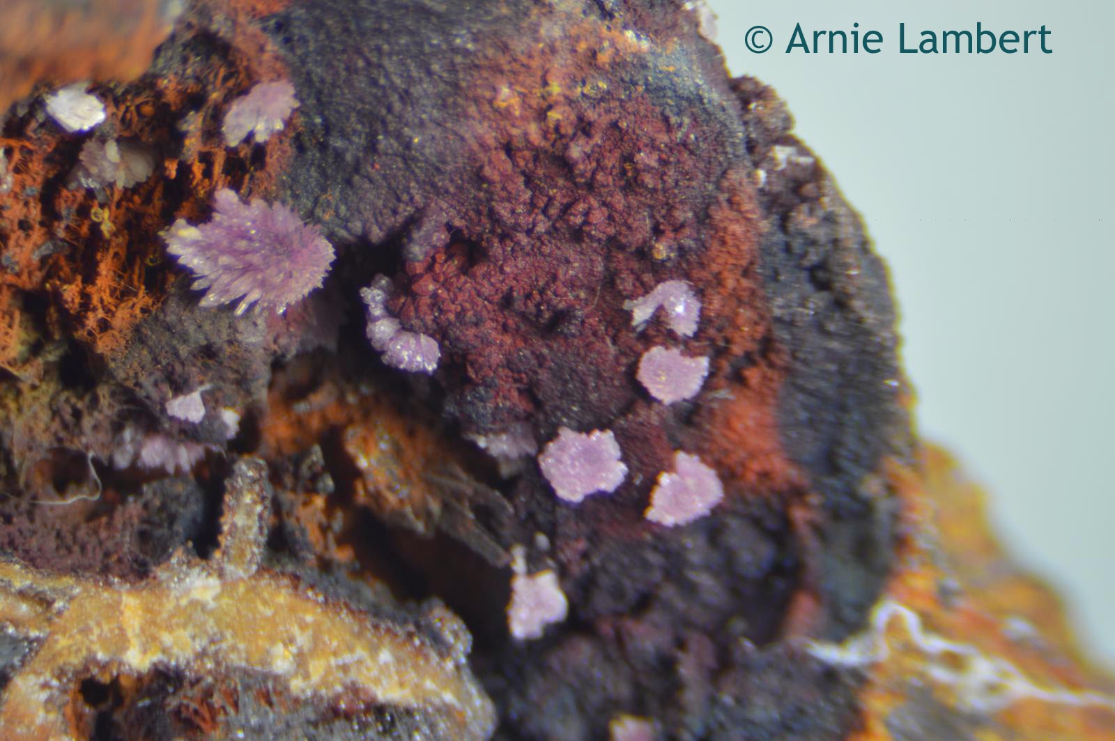
Strengite from Alabama under the microscope
Strengite from Indian Mountain in Cherokee Co., Alabama.
Strengite minerals as seen under the microscope.
Strengite from Indian Mountain in Cherokee Co., Alabama.
Thank you Arnie for sharing your images with Microscope World!
Thursday, September 18, 2014

Funghi Spores under the Microscope

A spore is a unit of asexual reproduction, often a part of the survival process. Spores can typically survive in unfavorable conditions and help form part of the life cycle of many plants including funghi. Spores are typically unicellular.

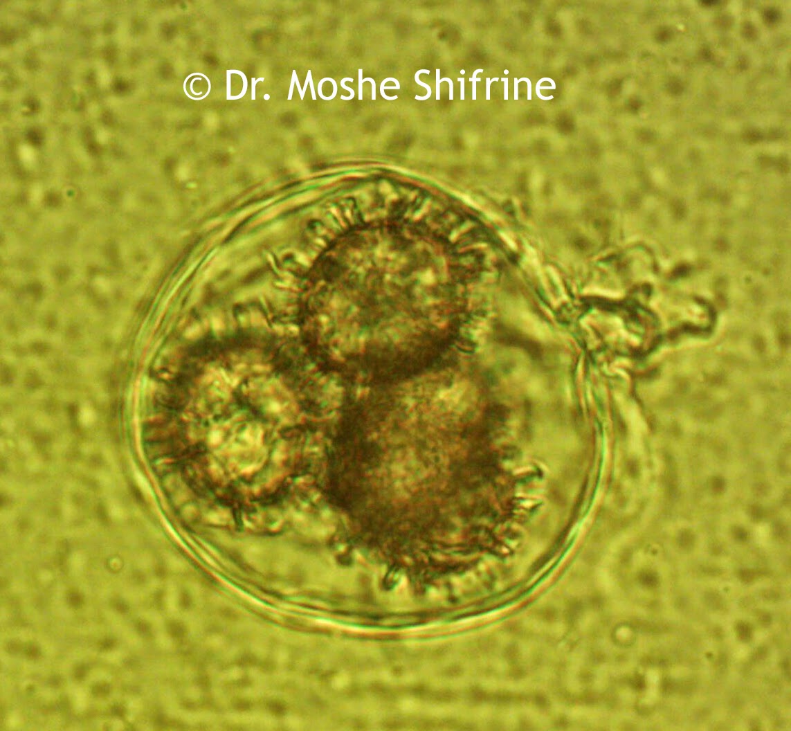
These funghi spore images were captured by Dr. Moshe Shifrine using the U2LCD digital tablet lab microscope. Dr. Shifrine is a retired Professor of Immunology (University of California at Davis) with a Ph.D. in microbiology specializing in mycology, the study of fungi or mushrooms.

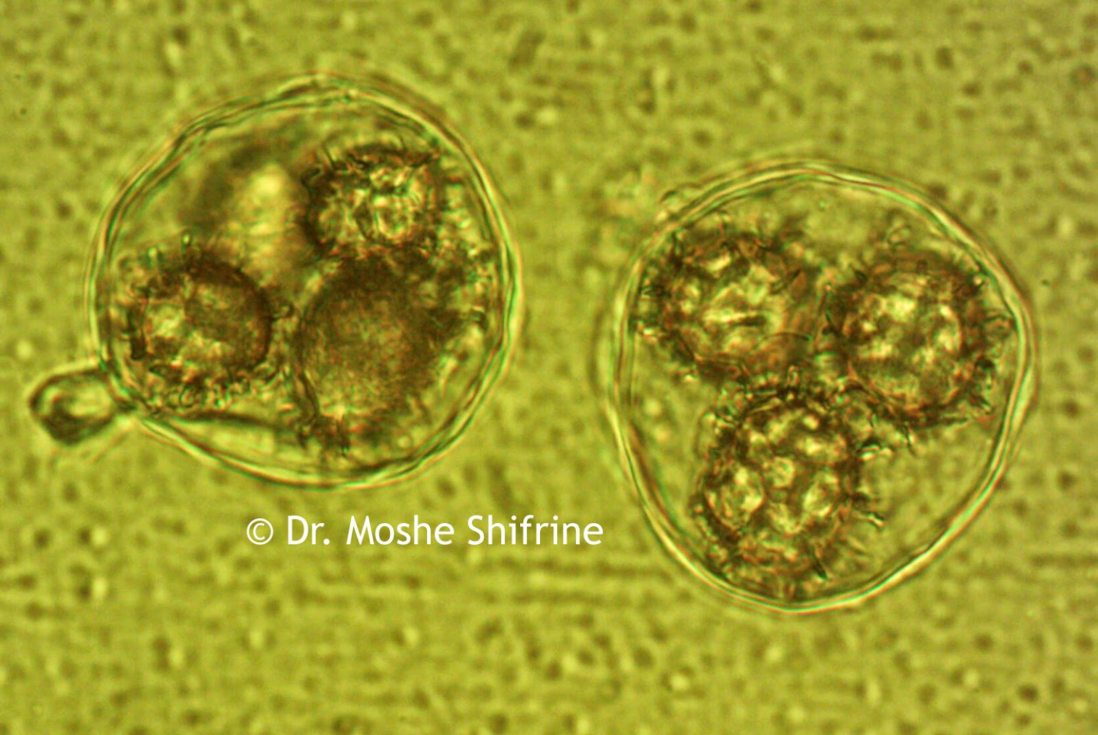
Funghi spore captured under a biological microscope at 1000x magnification.
Funghi Spore under the microscope at 1000x magnification.

Funghi spore, 1000x, under biological microscope.
Funghi Spore captured under the microscope at 1000x magnification.
Funghi spore 1000x magnification.
Funghi spore under the microscope at 1000x.

Friday, September 5, 2014

Sarcina under the Microscope

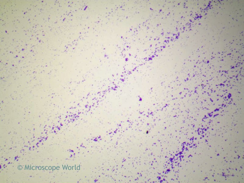
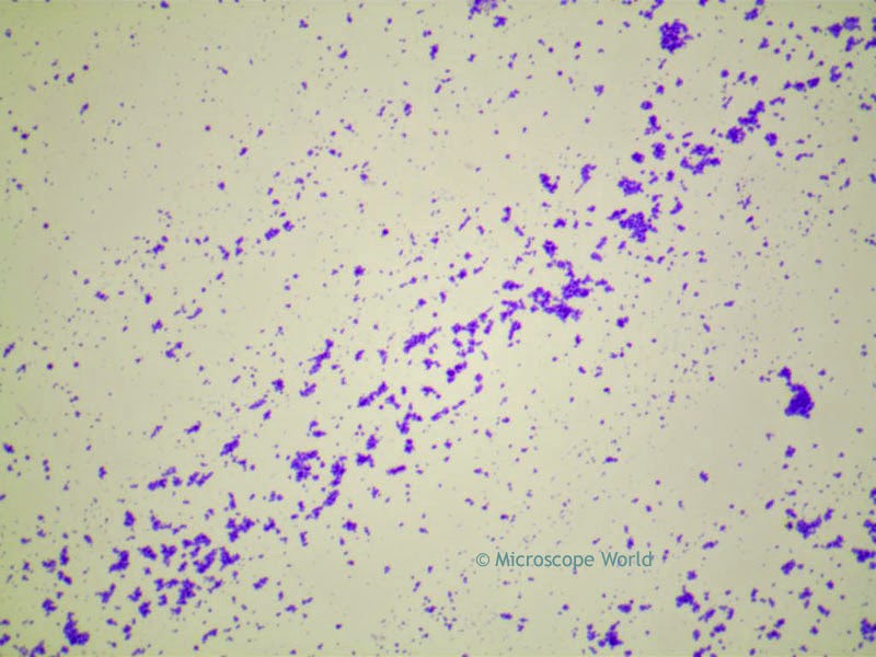
Sarcina is a genus of bacteria found most commonly in organic fluids in the stomach. The Sarcina prepared microscope slide is part of the Bacteriology Prepared Microscope Slide Kit.

Sarcina captured under the microscope at 40x magnification.
Sarcina captured under a biological microscope at 100x magnification.
Sarcina captured under the microscope at 100x magnification.
Sarcina captured under a biological microscope at 400x magnification.
All microscopy images were captured using the Richter Optica U2 biological microscope and a microscope camera with software.