Wednesday, April 30, 2014

Articulated Arm Stereo Zoom Microscope

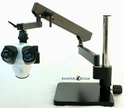
A new addition to the Richter Optica S6 series of stereo zoom microscopes is the S6-AASQ articulated arm microscope.

articulated arm stereo microscope
Richter Optica S6-AASQ Stereo Zoom Microscope

The S6 articulated arm stereo zoom microscope is affordable and provides the following:
  • Zoom range of 7x-46x.
  • 5-Year Warranty.
  • Optional LED ring light or LED dual pipe light available.
  • Available in binocular or trinocular for mounting a microscope camera.
  • High quality optics

Monday, April 28, 2014

Microscope Infinity Corrected Optics versus Fixed Tube Length

When microscopes were first introduced, all objectives had a fixed tube length. This means that there is a set distance from the nosepiece where the objective is screwed in, to the point where the ocular sits in the eyepiece. During the 19th century The Royal Microscopy Society standardized the microscope tube length to 160mm.

Using a tube length of 160mm works well, unless optical accessories such as a vertical illuminator or polarizing accessories are added into the light path of a fixed tube length microscope. If items are added, it changes that tube length to more than 160mm.  In order to solve this problem, in the 1980s the Infinity Corrected Optical System became common place.

Microscopes that utilize the Infinity Corrected Optical system have an image distance that is set to infinity. A tube lens is placed within the body tube between the objective and the eyepieces to produce the intermediate image (see image below). The Infinity Optical System allows auxiliary components such as illuminators, polarizers, etc. to be added into the parallel optical path between the objective and the tube lens with only minimal effect on focus.
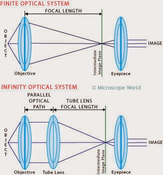
microscope tube length
Fixed tube length versus Infinity Corrected Optics
The Infinity Corrected Optical System typically makes it easier to maintain parfocality between different microscope objectives, even when auxiliary components are added to the microscope.

Friday, April 25, 2014

Bee Under Microscope

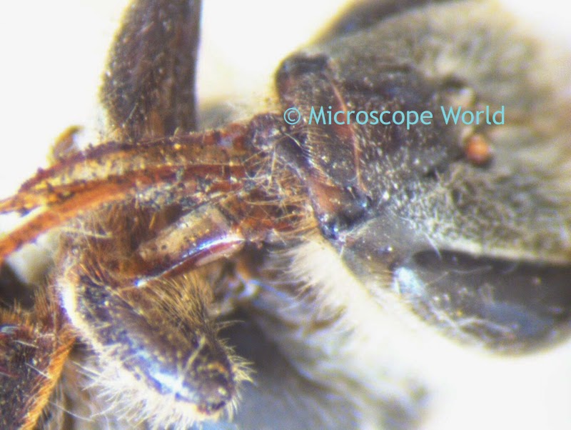
This bee was captured using the Richter Optica S6-D digital stereo zoom microscope. The image was captured at both 10x and 30x magnification.

Bee 10x under stereo microscope
Bee under the stereo microscope at 10x.
bee captured under microscope 30x
Bee under the stereo microscope at 30x.

Wednesday, April 23, 2014

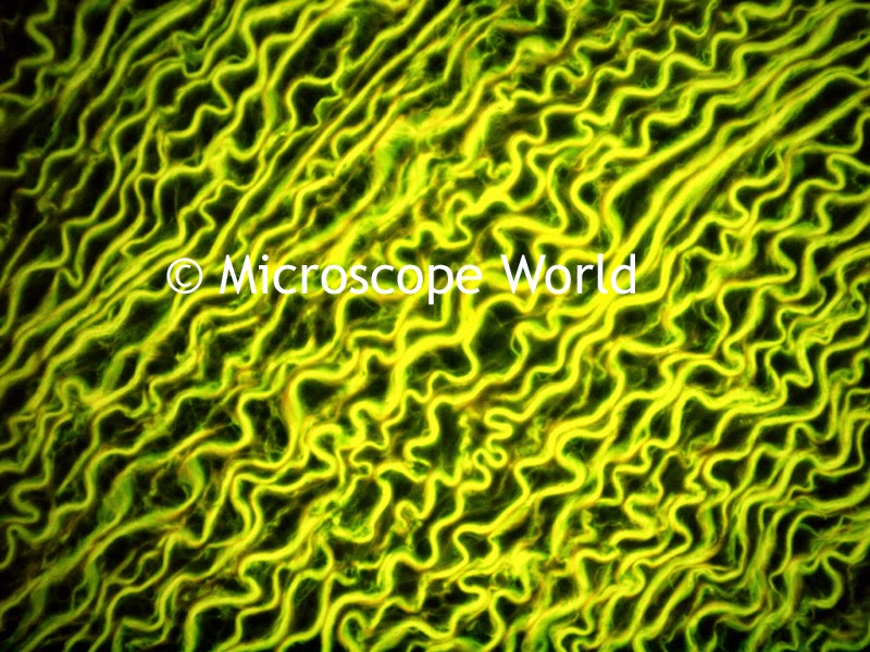
Aorta under the Microscope

The aorta is the largest artery in the human body. The aorta starts at the left ventricle of the heart and stretches down to the abdomen where it bifurcates into two smaller arteries. The aorta's function is to deliver oxygenated blood to various parts of the body through the systemic circulation.

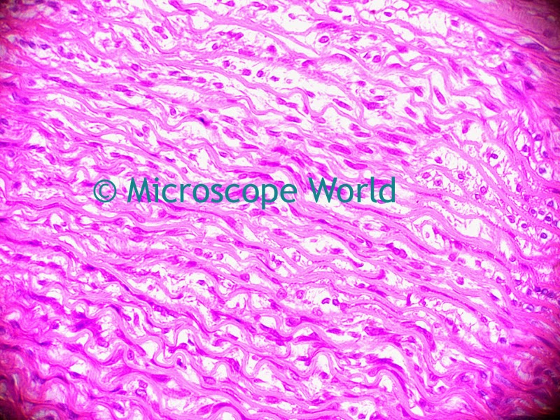
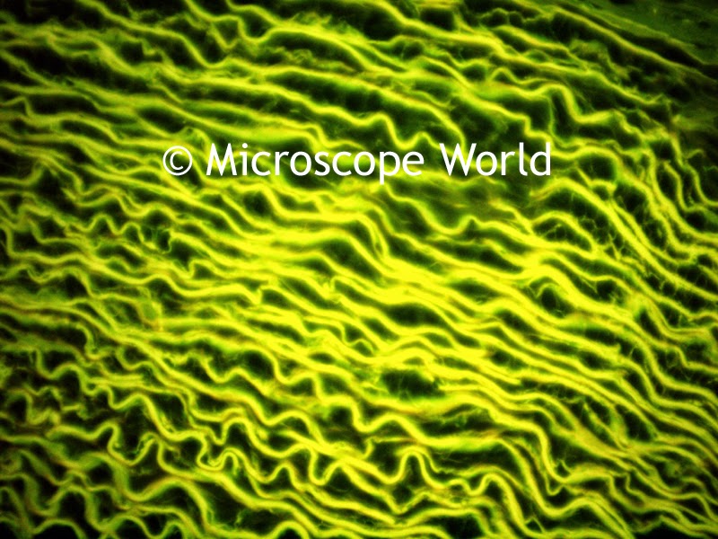
The microscope images below were captured first with brightfield and then with fluorescence microscopy.

microscope image of aorta, 400x
Aorta, 400x, captured with brightfield using the U2LCD microscope.
Fluorescence microscopy image 400x
Aorta, 400x, captured with the fluorescence microscope.
human aorta
Aorta Illustration courtesy Edoarado.
Microscopy images were captured using either the LCD tablet camera or the 5mega pixel microscope camera.

brightfield microscopy image
Human Aorta, 400x, Brightfield Microscopy

Human Aorta Fluorescence Microscopy
Human Aorta, 400x, Fluorescence Microscopy

Monday, April 21, 2014

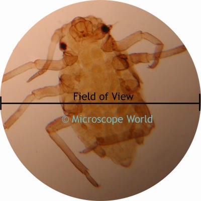
How to Calculate Microscope Field of View

The microscope field of view, or field diameter, is the distance across the image as seen through the microscope.  The field of view is greater at lower magnifications. For example, at 10x magnification 20mm might fill your field of view, whereas at 40x magnification you might only see 5mm.

microscope field of view image

Calculating field of view is a simple mathematical formula. You will need to know the field number of the eyepieces being used, the objective value, and any auxilary objective values.

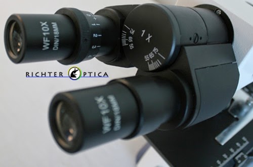
binocular microscope image
Notice the field number written on the eyepieces says 18mm.
Field Size = Field Number ÷ Objective Lens ÷ Auxiliary Lens

If you are using a stereo microscope with a 0.5x auxiliary lens on it, you would first need to look at the eyepiece to see what the Field Number (FN) is. If you were using 10x eyepieces and the field number was 20, and you wanted to find the field of view at 30x magnification, do you know what the math equation would look like?

When using 10x eyepieces, and a 0.5x objective, in order to have 30x magnification, the magnification knob would need to be set at 6x. The field of view formula is below:

Field Size = 20 (FN) ÷ 6 ÷ 0.5
Field Size = 6.67mm
At 30x magnification the entire field of view in the microscope setup mentioned above is 6.67mm.

Wednesday, April 16, 2014

Easter Science Projects!

This weekend is Easter and you might have some extra eggs at home. Here are a few science projects you can do at home with an egg!

Bouncy Egg:


Crystal Egg Geodes:

Learn how to make beautiful crystal egg geodes here.

Microwave Growing Peeps:
 
Take a peep, place it on a graham cracker and put it in the microwave for instant s'mores! Watch your peep get HUGE! Be careful though - much time over 10 seconds and your peep will burn.

Look at your science projects under the Microscope!

kids microscope
Kids Microscope 20x Magnification
What does the yolk of an egg look like under the microscope? How about the crystal geodes, or the peep, both before and after you made him grow in the microwave!
Happy Easter!!
 

Monday, April 14, 2014

Moth Larvae under the Microscope

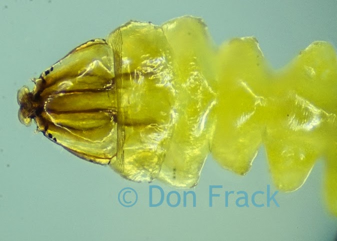
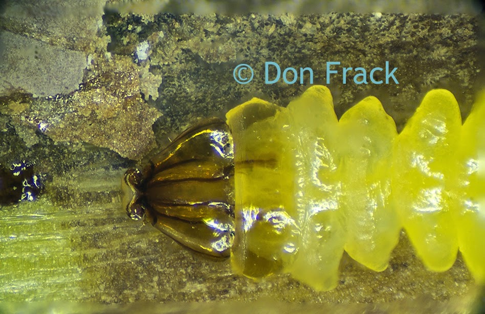
Don Frack is an entomologist and a customer of Microscope World. He recently captured these images of moth larvae at 45x magnification using the Meiji EMZ-5TRD stereo microscope, the PLS-3 transmitted LED stand, and LEDR-4 Ring Light System.

Moth Larvae under the microscope
Moth Larvae, 45x, captured with Pentax SLR camera.

moth larvae under stereo microscope
Moth Larvae, 45x magnification.

Tuesday, April 8, 2014

Tendons under Microscope

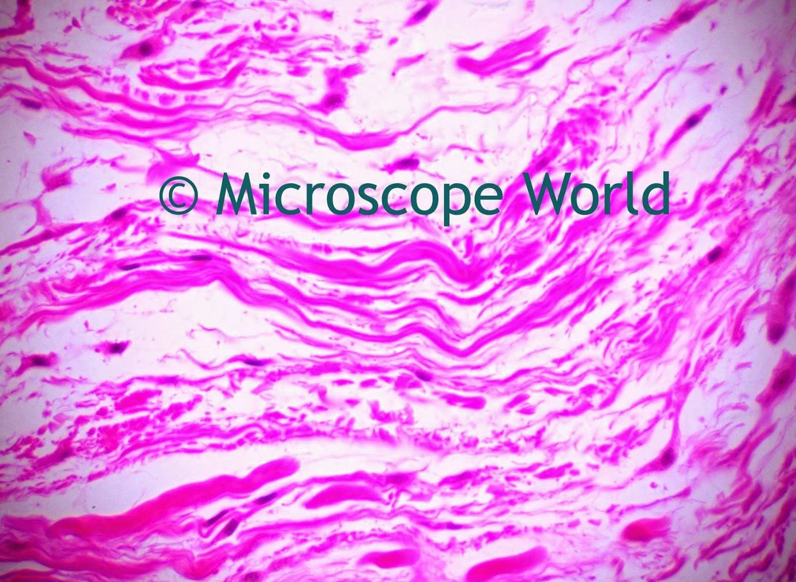
The human tendon is a tough band of fibrous tissue that connects muscle to bone. Tendons and muscles work together to move bones.

These images of human tendons were captured under the Richter Optica biological microscopes: U1 and U2LCD.

Tendons captured under microscope
Tendon captured at 100x with U1 Biological Microscope


The tendon prepared slide is part of the Human Histology: Human Organ Prepared Slide Kit.

Tendon at 400x
Tendon captured at 400x with the U2LCD digital microscope.

Friday, April 4, 2014

ESD Safe Microscopes

Electrostatic discharge (ESD) is the sudden flow of electricity between two electrically charged objects caused by contact, an electrical short, or dielectric breakdown. The ESD occurs when differently-charged objects are brought close together or when the dielectric between them breaks down, often creating a visible spark. Lightning is actually a large-scale ESD event.

ESD can damage sensitive electronic devices and therefore specifically in manufacturing of printed circuit boards or other quality control of electronics, ESD safe microscopes are required. Electronics manufacturers establish electrostatic protective areas free of static, using measures to prevent charging. ESD Safe stereo microscopes are used in these manufacturing environments.

ESD safe microscope
Motic SMZ-171 ESD Safe Stereo Zoom Microscope

Thursday, April 3, 2014

Blood under Microscope

This image is of human blood captured under the BA210LCD microscope at 400x magnification using brightfield.

Human blood under microscope 400x
Human Blood, 400x
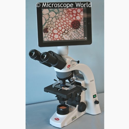
Motic BA210 lab microscope with LCD
Motic BA210 LCD Digital Microscope
The LCD microscope tablet camera on the BA210 microscope is an Android based touch screen tablet that allows the user to view and capture live images or video. The tablet is WiFi enabled and images can be emailed or saved to a micro SD card.

Wednesday, April 2, 2014

Brightfield versus Fluorescence: Columnar Epith

The images below were captured with a biological microscope using both brightfield and then with fluorescence. The prepared slides are a part of the Histology: Human Organ prepared slide kit.

microscope image columnar ciliated epith
Columnar Ciliated Epith captured at 400x with a brightfield microscope.

Fluorescence microscope image
Columnar Ciliated Epith captured at 400x under Fluorescence Microscope

microscope image at 400x
Pseudo Stratified Columnar Ciliated Epith, 400x, Brightfield Microscopy
fluorescence microscopy 400x image
Pseudo Stratified Columnar Ciliated Epith, 400x, Fluorescence Microscopy