Friday, June 29, 2012

Cinnamon Under Microscope

Cinnamon is a spice obtained from the inner bark of nearly a dozen trees found in the genus Cinnamomum. Cinnamon is used to flavor both sweet and savory dishes. Originally native of the island of Sri Lanka, cinnamon trees can now be found as well in South East Asia.

Cinnamon image courtesy of Luc Viatour.


Cinnamon is harvested by growing the tree for two years then coppicing it. The next year, about a dozen shoots will form from the roots.

Cinnamon stick captured at 10x magnification under the EMZ5TR-PBH stereo microscope using the MW5.1 CCD microscope camera.

25x Magnification.

45x Magnification.

Wednesday, June 27, 2012

Sunflower Moth Pupa under Microscope

Microscope World had a customer several years ago in the agriculture field. This particular customer was looking at moth pupa in sunflowers. A pupa is the life stage of some insects that undergo transformation. These insects go through four stages including: embryo, larva, pupa and imago (adulthood). Pupae are inactive and can not usually move around. They typically have a hard protective covering and are often camouflaged to prevent predators from eating them.

Moth pupa resting inside a sunflower stem - you can see it just above the word "Microscope".

This is the head of the pupa, captured with 80x magnification using a stereo microscope and a consumer SLR digital camera, and a digital camera adapter.

Thursday, June 21, 2012

Mushrooms under the Microscope

This is a simple and fun kids science project. Next time you are at the grocery store, pick out a large porotabello mushroom.

On the under-side of the mushroom remove several of the "gills" and place them under a stereo low power microscope. What do you notice about the patterns in the gills?

A mushroom is a fleshy, spore-bearing fruiting body of a fungus. Even the mushrooms that are safe to eat are considered fungus! The mushrooms purchased in the grocery store are safe to eat, but do not eat mushrooms found in the wild - many are quite poisonous and will make you sick.

Next, take a small thin section of the underside of the mushroom and place it on a slide under a high power biological microscope. (Make sure the section is thin enough to allow light to pass through it). What differences do you notice at higher magnification?

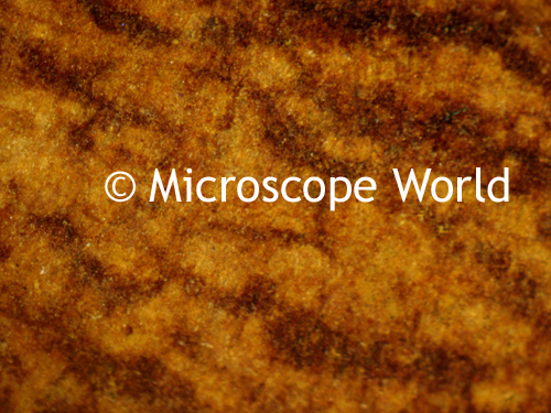
Outside, take a look around some large trees that receive plenty of water in the summer. Do you see any growth or fungus on the tree such as the one shown below?

Once you locate some tree growth or fungus, remove a small piece from the tree and look at it under your stereo microscope. Then slice a small thin piece to view under the compound microscope. What do you think makes up the variety of colors in the fungus?

In order to view spores from the mushroom, take a fresh mushroom purchased from the store and cut the stem off. Place it cap-side down (gills on the bottom) on a blank glass slide and cover it with a bowl overnight. Some spores should fall off the mushroom in the night and end up on the slide. Carefully remove the mushroom and cover the slide with a cover slip. Using a compound biological microscope look at the slide. What do you notice? Are the spores uniform in size? Document your findings and share them with your science class when you return to school in the fall.

Friday, June 15, 2012

Polarizing Microscope Characteristics

Polarized light microscopy is often utilized by geologists to study naturally occurring minerals and rocks in thin sections, and by mineralogists and ceramicists for research in industrial environments. A polarizing microscope is also heavily used by scientists who study the various phase transitions and textures exhibited by liquid crystalline compounds, and polymer technologists often make significant use of information provided by the polarized light microscope. Forensic scientists take advantage of polarized techniques in the analysis of fibers, hairs, and other particles that are discovered at crime scenes. The advantages of polarized light have been utilized to explore biological processes.

All polarizing microscopes utilize a polarizer and analyzer in conjunction with each other. Polarizing microscopes also include strain-free objective lenses and condenser, a Bertrand lens, a circular graduated stage capable of 360-degree rotation, and an opening in the microscope body or intermediate tube for compensators, such as a full-wave retardation plate, quartz wedge, or quarter-wavelength plate. If you were to remove the polarizer and analyzer (while keeping other components in place) from the light path, the instrument becomes equal to a typical brightfield microscope with respect to the optical characteristics.


Other than the polarizer and analyzer making up an essential part of a polarizing microscope, these items are typically found on a polarizing microscope:
  • Strain Free Objectives: Stress introduced into the glass of an objective during assembly can produce unwanted optical effects under polarized light, compromising performance. Objectives designed for polarized light observation are distinguished from ordinary objectives with the inscription P, PO, or Pol on the barrel. 
  • Polarizing Condenser: The polarizing microscope condenser includes a built-in rotatable polarizer and a strain free optical system, just like the objective lenses.
  • Polarizing Rotating Stage: The 360-degree circular rotating specimen stage facilitates orientation so that the objectives can be centered with the stage and the specimen's center field of view. Most polarized microscope stages will include a vernier scale so rotation angle can be measured to an accuracy of 0.1 degrees.
  • Bertrand Lens: This specialized lens projects an interference pattern formed at the objective rear focal plane into focus at the microscope image plane. The lens is designed to enable easy examination of the objective rear focal plane, to allow accurate adjustment of the illuminating aperture diaphragm and to view interference figures. The bertrand lens is located between the analyzer and eyepiece for easy positioning in and out of the light path.
  • Compensators and Retardation Plates: Many polarized light microscopes contain a slot allowing for the insertion of compensators and/or retardation plates between the cross polarizers, which are used to enhance optical path differences in the specimen.
  • Quartz Wedge: A quartz is used to measure retardation and determining the Z direction.
Polarized light microscopy can use both reflected and transmitted light. Reflected light is useful for the study of opaque materials including ceramics, mineral oxides and sulfides, alloys, metals, composites and silicon wafers. Reflected light techniques require a dedicated set of objectives that have not been corrected for viewing through the cover glass. View images captured with a polarizing microscope here.

Wednesday, June 6, 2012

Jewelry Microscope

Viewing gems and precious stones under a microscope can sometimes prove tricky based on the reflective nature of cut stones. A gemological microscope is usually best with a gem clamp and a darkfield attachment.

These jewelry images were captured with a Meiji gemological microscope using a microscope c-mount adapter with the MW5.1 CCD microscope camera and included software.


Most gemological images are captured at a low magnification, typically between 10x - 20x.

Monday, June 4, 2012

Stereo Microscope and Fibers

A customer of Microscope World recently needed to inspect the fibers they manufacture. The system they used included a stereo zoom microscope on a lighted stand. The images were captured with a microscope camera and the included software.

Fibers captured at 10x magnification with a top light using the clear glass stage plate.

Fibers captured at 30x magnification using the bottom illumination only along with a blue filter.

Fibers captured at 40x magnification under a stereo microscope with top light illumination.

Coarse fibers captured with 60x magnification.

Four single fiber ends captured at 125x magnification.