Tuesday, May 29, 2012

Muscovite (Mica)

Muscovite (also known as mica) is a mineral made of aluminium and potassium. It is made up of very thin sheets which are often elastic. Sheets of muscovite that are huge (5 meters by 3 meters) have been discovered in Nellore India.

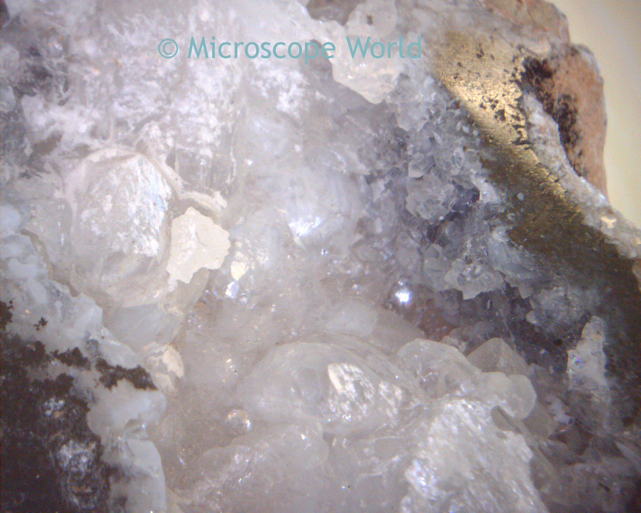
Muscovite with albite from Doce valley, Minas Gerais, Brazil. 
Photo: Rob Lavinsky

Muscovite can be colorless or tinted through grays, browns, greens, yellows or very rarely, reds. It is the most common mica, found in granites, pegmatites, gneisses, and schists. In pegmatites it is often found in immense sheets that are commercially valuable. It is in demand for fireproofing and insulating materials and to some extent as a lubricant.

Muscovite image captured with a polarizing microscope using the ProgRes C14+ microscope camera.

The name muscovite comes from Muscovy-glass, a name formerly used for the mineral because of its use for windows in Russia.

Thursday, May 24, 2012

Salty Liquorice

Salmiak (also known as salty liquorice) is a variety of liquorice flavored with ammonium chloride, common in Nordic countries, Netherlands, and Northern Germany. The ammonium chloride gives the liquorice a salty taste, which some describe as tongue-numbing.


Salmiak is usually an acquired taste and often people not familiar with the strong taste find it overwhelming. The candies are almost always black and can range from very brittle to soft in texture. The flavor of salty liquorice is sometimes used in ice cream or alcoholic beverages.

This microscope image of salmiak was captured with a Jenoptik ProgRes microscope camera.

Tuesday, May 22, 2012

Quartz & Metal Under Microscope

Quartz and metal can both have reflective traits that sometimes make them difficult to view under the microscope. A simple solution is an inexpensive handheld camera, which provides a small amount of magnification.

The MW1-LD2 works well for these basic applications. This camera connects directly with an included USB cable into the computer and includes software for capturing images, making measurements and labeling images. Typically the ambient light from a well lit room is plenty to illuminate the object.

Image of quartz captured with the MW1-LD2 digital camera.

Metal captured with the MW1-LD2 camera.


Monday, May 14, 2012

Aphids and Digital Microscopes

The MW2-HD1 digital high school microscope provides the ability to view and capture images on the computer, with quality educational optics, at an affordable price.

Aphids are small, sap-sucking insects, sometimes referred to as plant lice. They can be some of the most destructive insects. From a zoological standpoint, the aphid is a very successful insect!

Aphids are popular in temperate zones and surprisingly don't do well in tropical areas.


Tuesday, May 8, 2012

Electronics Inspection Microscopes

Electronics inspection microscopes combine high quality optics with rugged dependable microscope construction so that printed circuit board manufacturers can problem solve during production.


Printed circuit board captured at 70x magnification.

Printed circuit board captured with C14+ microscope camera.

Electronics inspection microscopes typically provide magnification in the range of 5x-25x. Although occasionally an auxiliary lens will be added to provide higher magnification (shown in the image above at 70x magnification).

Friday, May 4, 2012

Fluid Channel under Microscope

Fluid channel for a press button. Captured using a Jenoptik ProgRes microscope camera. Image courtesy of HSG-IMIT Microtechnology.

Thursday, May 3, 2012

Brain Research

Dr. Axel Schleicher captured this cross section image of the brain while working for the Institute for Brain Research, University of Dusseldorf. Using a biological microscope and a ProgRes camera for excellent color reproduction and high resolution, this image was captured.

Front cut through the brain hemisphere of a Rhesus-Ape-Immune-histochemical presentation of NO-receptive Guanylylcyclasis.