
Darkfield illumination is a technique where the direct light path is blocked and only oblique light rays are permitted to hit the specimen. This results in an image that appears to almost be back-lit.
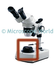 An example of a stereo microscope darkfield attachment on the National Optical stereo microscope.
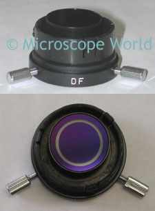 This is the high power darkfield attachment for the National Optical 162 biological microscope. This is a simple plug-in type of darkfield attachment that replaces the condenser.
Darkfield microscopy is often used when viewing blood cells, gems, cell membranes and tissue.