Monday, March 31, 2014

Introducing Swift WiFi Microscopes!

Swift-Microscopeworld.com is excited to offer two new Swift WiFi microscopes:
The Swift WiFi microscopes create their own wireless network that any Android or iOS device can connect to. The microscope also has on-board software that can be controlled with a web browser on a computer for image capture and measurement.

WiFi microscope from Swift
Swift WiFi Microscope M3702C-4DGLX

Students can use their smart phone, tablet or computer to view the images seen under the WiFi microscope. Streaming images are sent to up to 6 devices without the need for a router. The Swift WiFi microscope generates its own WiFi signal and can be used separate from your existing network. You can download the free MotiConnect App at the Apple App store or at the Google Play store.

The biological digital WiFi microscope includes a built-in 1.3 mega pixel camera and allows capture of both live images and video. View the Swift WiFi microscope here.

Friday, March 28, 2014

Dicot under the Microscope

These images were taken of the Dicot prepared slide (part of the Microscope Starter Kit) using a Canon digital SLR camera adapter on the Motic BA310 microscope. All images were captured at 400x magnification.

Dicot under the microscope
Dicot, 400x under biological microscope.

Dicot 400x magnification
Dicot, 400x under biological microscope.

Dicot at 400x image
Dicot, 400x under biological microscope.

Dicot under the microscope
Dicot, 400x magnification.

Wednesday, March 26, 2014

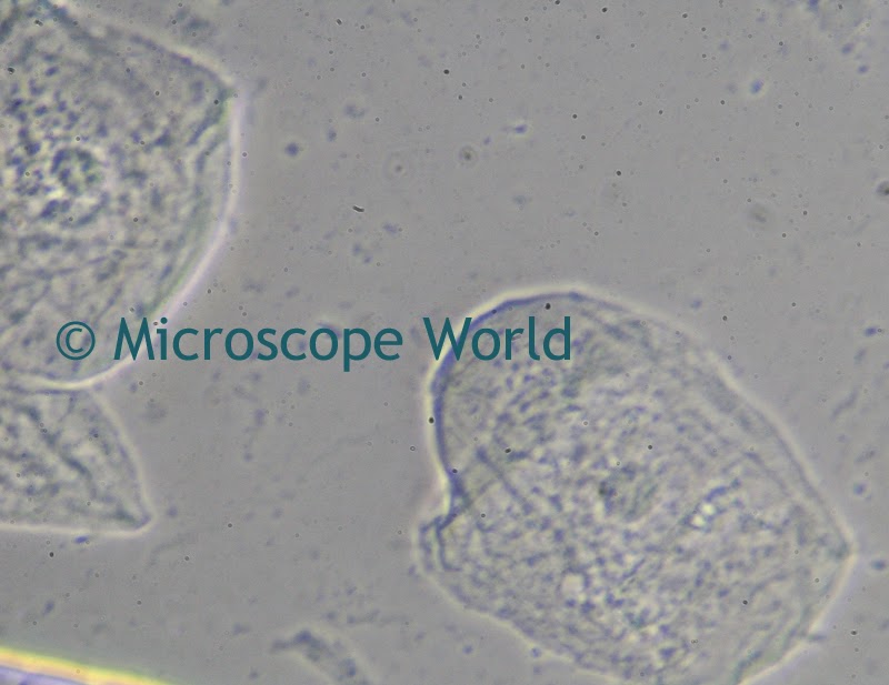
Cheek Cells under the Microscope

Cheek cells are one of the easiest cells to view under a microscope, since everyone comes equipped with them! Take a Q-Tip and swab the inside of your cheek. Rub the moist Q-Tip on a blank glass microscope slide and place a cover slip on top.

Using a biological microscope, view the slide at the lower magnification first and then move up to higher magnifications. If you have access to a phase contrast microscope you will be able to view even more detail in the cheek cells.

The following images were captured using the U2 biological microscope and a 40x Phase Contrast Objective lens and phase contrast condenser.

microscope cheek cells 40x

image microscope cheek cells

cheek cells under phase contrast microscope

phase contrast cheek cells image

microscope image

microscopy cheek cells image

Monday, March 24, 2014

High School Microscope Features

The Richter Optica HS-1+1 is a popular high school microscope for several reasons.

student microscope labeled diagram
HS-1+1 Student Microscope
  • Separate Coarse & Fine focusing knobs are important. Without a fine focusing knob, it is very hard to get a crisp and clear image at 400x or higher magnification.
  • A head that rotates makes it easier to position students around the microscope.
  • The 2nd eyetube allows for the addition of a microscope camera, or multiple students can view specimens at the same time.
  • The 5-position disc diaphragm helps adjust the amount of light that passes through the specimen and will allow for crisp images.
  • The rheostat control also allows for light control. Too much light can result in washed-out images.
  • LED lighting is cool and will not harm living specimens.
The video below demonstrates the features on the HS-1+1 student microscope.


Thursday, March 20, 2014

C. Elegans under the Microscope

Caenorhabditis Elegans (C. Elegans) is a nematode and a member of the phylum Nematoda. C. Elegans are roundworms and threadworms that are smooth-skinned, unsegmented worms with a long cylindrical body shape tapered at the ends. They are non-hazardous, non-infectious and live in the soil. The worms are often found in soil near rotting vegetation, feeding on microbes such as bacteria.

Scientists often study C. Elegans because they are about as primitive an organism that exists which shares many of the essential biological characteristics that are central problems in human biology. The worms have a nervous system with a brain and exhibit behavior that is capable of rudimentary learning.

Maximum length is about 1mm long and when studied, the worms are typically grown on petri dishes seeded with bacteria. All 959 somatic cells of the C. Elegan's transparent body are visible with a microscope and the average life span is only 2-3 weeks.


The video above was captured using the Lumenera 2-1 microscope camera and a stereo fluorescence microscope.
C. Elegans
Illustration courtesy (KDS444) Wikipedia

Monday, March 17, 2014

What exactly are Stereo Zoom Microscopes?

Stereo zoom microscopes provide lower magnification and reflected light with two eyepieces for viewing a "stereo" image. The eyepiece magnification combined with the objective lens magnification provides the total overall magnification, and creates a visual effect of depth of perception for the viewer. This three-dimensional capability produces laterally precise, highly detailed images, offers a larger field of view, and allows for extended working distances making stereo zoom microscopes ideal for viewing larger objects in lab, industrial and educational settings.

These microscopes are often used in jewelry and watchmaking, circuit board inspection, dissection, microsurgery, and forensic engineering because they are ideal for observing large specimens and analyzing materials with complex surfaces.

Stereo zoom microscopes are available in two configurations:
  • Greenough design: the most commonly used stereo microscope. Each eye has a separate optical path and the angle of divergence creates a shallow "V" design, which results in a three dimensional image.  The greenough design is typically less expensive, and is best for examining and inspecting specimens at lower magnification of anywhere from 5x-90x.
  • Common Main Objective design: for higher magnifications, the common main objective stereo microscope provides a better image. These microscopes are more popular for research and development because of the higher resolution images that they produce. The common main objective stereo microscope has a parallel optical path and at magnifications above 90x images are much more crisp and clean.
Stereo Microscope image
S6 Stereo Microscope (Greenough Design)

Stereo microscopes are available with either a binocular (two eyepieces) or trinocular (two eyepieces plus a camera port) head. The trinocular port typically has a beam splitter on the head of the microscope which when engaged, will allow light to be directed up to the camera and away from the eyepieces, resulting in higher quality captured images. Microscope cameras are great tools for classroom study, research documentation or creating reports.

Huvitz HSZ-700 Trinocular Microscope
Huvitz HSZ7T-PL Trinocular Microscope (Common Main Objective)

If you have questions about stereo zoom microscopes, or would like to speak with a specialist, please contact us today.

Friday, March 14, 2014

Pollen Under the Microscope

Pollen can be seen under the microscope best at about 400x magnification. Pollen is the powder that often falls off flower stamens if touched. The bee shown below has yellow pollen on him.

bee with pollen image
Photo courtesy: André Karwath
The images below of pollen were captured using the U1B biological high school microscope and a 2 megapixel microscope camera.

pollen 100x under microscope
Pollen captured at 100x with U1B microscope.
400x microscope pollen image
Pollen captured at 400x with U1B microscope.

Wednesday, March 12, 2014

Microscope Mechanical Stage

What exactly is a microscope mechanical stage, and do you really need one? A biological microscope that does not have a mechanical stage on it will look like the image shown below with two stage clips for holding the microscope slide in place. A microscope mechanical stage is not required in order to view images, but it makes using the microscope much easier and less frustrating at higher magnifications.

microscope stage image
Microscope stage with stage clips (no mechanical stage).
A mechanical stage holds the microscope slide in place and gives the user much more control over the slide. At very high magnifications (400x or 1000x) when you want to move the slide just slightly to view a different part of the specimen, if you are not using a mechanical stage you must move the slide with your fingers. It can be very easy to push the sample right out of the field of view.

When using a mechanical stage one of two knobs are rotated to move the slide in very small increments either left to right or forward and back. The microscope mechanical stage below can be put on the microscope above by removing the stage clips and screwing the mechanical stage onto the flat microscope stage.

microscope mechanical stage
Microscope Mechanical Stage
Below is a video demonstrating the Richter Optica U2 microscope which has a built-in mechanical stage with low-position drop-down controls for controlling the stage.

)

If you have questions about adding a mechanical stage to your microscope please contact Microscope World.

Saturday, March 8, 2014

Industrial Microscopes in the Automation Industry

Industrial microscopes are being used in the automation industry to speed up inspection, analysis, and quality assurance processes for a broad range of objects and surfaces. In one fully-automated example, a six-axis robotic arm "feeds" samples to a variety of industrial microscopes in a sequence so that all systems can be used simultaneously. This in turn produces measurements and high resolution imagery of both finished and incomplete products that may not pass crucial inspection before moving to the next step in the process.

In other examples, microscopes are automated to produce high resolution images of pathology samples for molecular diagnostics and cytometry, enhancing and automating the workflow of investigators in the pathology laboratory. In some labs, automated microscopes are used to capture live-cell imagery and are especially useful for acquiring images in time-lapse experiments over set periods of time.

Using industrial microscopes in the automation industry, you can benefit from:
  • Reproducible methods for exact positioning of specimens.
  • Automated processes that move the stage and focus on specimens.
  • Microscopy software can capture images for analysis or reporting.
  • Measuring items that will not fit within one field of view.
  • Switching from a variety of dedicated equipment to one multi-function tool for automated microscopy, digital imaging, measurements, image analysis and reporting.
  • produce reports from high-resolution samples.
  • High-resolution overviews for the inspection process.
Automated microscopy can sometimes allow you to trade in your optical comparator, metrology inspection station, toolmaker's microscope, or dedicated image processing station for an automated industrial microscope that can do the job of several tools as well as several individuals.

Thursday, March 6, 2014

Tripneustes Ventricosus Larvae under Microscope

A customer of Microscope World's, Mr. Martin Moe, rears both Diadema and Tripneustes. This is a photo he captured of a Tripneustes Ventricosus Larvae at day 32 with posterior pedicellaria and cilia ridge just before settlement.

Tripneustes Ventricosus (commonly called the West Indian sea egg), is a species of sea urchin. These urchins are found commonly in the Caribbean Sea, Bahamas, and Florida at depths of less than 33 feet.

Tripneustes Ventricosus feeds on algae, but tends to avoid highly calcified coralline algae.

Tripneustes Ventricosus image
Tripneustes Ventricosus - image courtesy of Martin Moe.
The Tripneustes Ventricosus image above was captured using the Meiji MT4300 biological microscope.