Microscope information, images from beneath the microscope and educational science projects.
Tuesday, December 22, 2015
Wednesday, December 16, 2015
Beer Brewing & Wine Making Microscopes
Whether you are an expert home brewer, own a company brewing beer, or own a vineyard, a quality beer brewing or wine making microscope can make your job much easier. Here are a few things to look for when selecting a microscope.
Beer and wine microscopes vary from basic phase contrast and digital basic phase contrast to more advanced full phase contrast microscopes or even an advanced fluorescence beer/wine microscope in order to perform live-dead tests.
Beer Brewing Microscope Tips:
- You will need magnification in the 400x to 1000x range in order to view cells, bacteria and yeast. If you simply want to count cells, a basic binocular compound microscope such as the basic beer and wine microscope with 400x magnification can do the trick.
- Make sure you have coarse and fine focusing! The fine focus adjustment is important, especially when using the microscope at the higher magnifications.
- Get a microscope with a mechanical stage. The mechanical stage will allow you to maneuver your slide in small increments and without frustration.
- A monocular (single eyepiece) microscope will suffice, although if you spend much time looking through the microscope binocular (two eyepieces) will be much more comfortable.
- Don't use a microscope with a disc diaphragm - an iris diaphragm on the condenser will allow you to enhance the contrast in your image when you close down the diaphragm.
- Phase contrast is the best technique for looking at both yeast and bacteria. Phase contrast microscopes vary from simple phase contrast (usually only 40x phase objective) to full phase contrast which provides phase contrast 4x, 10x, 40x, 100x objectives. Phase contrast will allow you to view enhanced images of both yeast and bacteria.
- Cell counting can be performed with a grid eyepiece reticle or with measurement software included with the microscope digital camera. A hemocytometer is used for counting.
- The Brewing Science Institute has some great educational info here.
Beer and wine microscopes vary from basic phase contrast and digital basic phase contrast to more advanced full phase contrast microscopes or even an advanced fluorescence beer/wine microscope in order to perform live-dead tests.
 |
| Beer Brewing / Wine Making Microscope |
Wine Microscope Tips:
Here are a few tasks every vintner should be able to perform with a microscope.
- Distinguish between bacteria, yeast and fungi under the microscope.
- Identify and differentiate living organisms from plant debris, filter agents or crystals.
- Identify the most common organisms by sight in order to take action quickly if needed - including wine yeast, mold and bacteria. Phase contrast microscopes are helpful when viewing bacteria and yeast.
- Count yeast cells and distinguish between living and dead cells.
Tuesday, December 8, 2015
Wright's Stain for Microscopy
Wright's stain is a histologic stain that facilitates the differentiation of blood cell types. It is classically a mixture of eosin (red) and methylene blue dyes. It is used primarily to stain peripheral blood smears and bone marrow aspirates which are examined under a light microscope. In cytogenetics, it is used to stain chromosomes to facilitate diagnosis of syndromes and diseases.
Wright's stain is named for James Homer Wright, who devised the stain in 1902. The stain is actually a modification of the Romanowsky stain. Because Wright's stain distinguishes easily between blood cells, it became widely used for performing differential white blood cell counts, which are routinely ordered when infections are suspected.
The blood smear images below have Wright's stain applied to them and were captured with a laboratory microscope.
 |
| Blood smear with Wright's Stain captured at 40x under a clinical lab microscope. |
 |
| Blood smear with Wright's Stain captured at 100x under a clinical lab microscope. |
 |
| Blood smear with Wright's Stain captured at 400x under a clinical lab microscope. |
 |
| Blood smear with Wright's Stain captured at 400x under a clinical lab microscope using a Plan Fluor objective. |
Friday, December 4, 2015
Understanding Microscope Resolution
A common misconception among first-time microscope purchasers is that more magnification is always better. Unfortunately, any microscope that advertises magnification above 1000x is offering empty magnification. You can learn more about empty magnification here. The limiting factor in a microscope is not its magnification, but rather its resolution. Resolution refers to the shortest distance between two separate points in a microscope's field of view that can still be distinguished as distinct entities.
Imagine a drawing that was made with large children's crayons, versus a drawing made with a sharp pencil. Microscopes with poor resolution will provide images that are similar to the crayon drawing, as the images will not be crisp and clear.
Numerical aperture determines the resolving power of a microscope objective lens, but the total resolution of the entire microscope is also dependent on the numerical aperture of the condenser (located beneath the stage on a compound biological microscope). The higher the numerical aperture of the complete microscope system, the better the resolution will be.
Aligning the microscope optical system is also important to ensure maximum microscopy resolution. The condenser must match the objective with respect to numerical aperture and adjustment of the aperture iris diaphragm. There is a great article here that discusses how to properly align the condenser as well as set the field iris diaphragm. The sketch below shows alignment of the iris diaphragm on the condenser (3) to match the objective value (4) of the current objective being used.
 |
| Iris Diaphragm on Microscope Condenser |
Wavelength of light is an important factor in the resolution of a microscope. Shorter wavelengths will yield higher resolution. Therefore, the greatest resolving power in optical microscopy is realized with near-ultraviolet light which has the shortest effective imaging wavelength. When using near-ultraviolet light special Near-Ultraviolet microscope objectives are used.
The resolution of a microscope provides the ability to view samples and specimens clearly. The main factor in determining resolution is the objective numerical aperture, but resolution is also dependent upon the type of specimen, coherence of illumination, degree of aberration correction and other factors such as contrast enhancing methodology in either the microscope's optical system or the sample itself.
Contributing Source: MicroscopyU
Tuesday, November 24, 2015
Dissecting Microscopes
What exactly is a dissecting microscope? There are several different types of dissecting microscopes and we will cover each of them below. A dissection microscope is also referred to as a stereo microscope or a low power microscope.
 A few features common among all dissecting microscopes:
Single power dissecting microscopes offer one single magnification. For example, the microscope might provide 20x magnification. These single power instruments are common in museums or schools where only one magnification is required to view samples. With a single power magnification dissection microscope it is possible to have two magnifications when using a different pair of eyepieces. For example, with a 10x and 15x pair of eyepieces (when the built-in objective is 2x) total magnification would be 20x and 30x. This is an inexpensive way to achieve multiple magnifications from a lower cost microscope.
Dual power dissecting microscopes provide two set magnifications. By turning a turret on the bottom of the body of the microscope (the black portion on the image at right), it is possible to flip back and forth between the two magnifications. The objective lenses are built into the microscope (for example, 1x and 3x) and the eyepieces can be changed. On a microscope that includes 10x eyepieces with a built-in 1x and 3x objective, the total magnification is 10x and 30x. Magnification could be increased by purchasing a pair of 15x eyepieces, which would provide 15x and 45x magnification. These microscopes are sometimes used by coin collectors, in schools, and in industrial quality control settings where an inexpensive microscope is required.
Stereo zoom microscopes provide a magnification range and all magnifications within this range can be viewed. The S6 stereo microscope shown at left provides magnification of 6.7x - 45x, which means you can see 10x, 15x, 18x, etc. all the way up to 45x. These are the most commonly used dissecting microscopes, since they provide more options for viewing the exact part of the sample at the desired magnification. Typically when increasing magnification on a stereo zoom dissection microscope, rather than changing eyepieces, an auxiliary lens is added to the bottom of the stereo microscope body. If you were using the microscope shown at left and wanted a bit more magnification, you might add the 1.5x auxiliary lens, which would change the magnification range to 10x - 67.5x. Stereo zoom microscopes are typically used in universities, in industrial settings for quality control and to view small parts, and in manufacturing.
 A few features common among all dissecting microscopes:
- Dissecting microscopes have working room on the stage. There is room to place larger objects such as rocks or nuts and bolts. In order words, the microscopy sample does not need to be on a microscope slide.
- Dissecting microscopes use light from above the stage, making it possible to view opaque objects. Some dissecting microscopes also have light beneath the stage, but not always.
- Dissecting microscopes provide low magnification (usually 10x - 40x is a common range) and they are used to view detail in objects you can already see with the naked eye. Blood cells are not viewed with a dissecting microscope, but rather with a compound microscope.
Single Power Dissecting Microscopes
 |
| Single Magnification Microscope |
Single power dissecting microscopes offer one single magnification. For example, the microscope might provide 20x magnification. These single power instruments are common in museums or schools where only one magnification is required to view samples. With a single power magnification dissection microscope it is possible to have two magnifications when using a different pair of eyepieces. For example, with a 10x and 15x pair of eyepieces (when the built-in objective is 2x) total magnification would be 20x and 30x. This is an inexpensive way to achieve multiple magnifications from a lower cost microscope.
Dual Power Dissecting Microscopes
 |
| S2 Dual Magnification Microscope |
Zoom Dissecting Microscopes
 |
| Stereo Zoom Microscope |
Friday, November 20, 2015
Insects Under the Microscope
Microscope World recently wanted to compare two different microscopy cameras for quality and differences in color. The two microscope cameras included the DCM5 documentation camera (5 megapixels) and the HDCAM7 high definition camera (8 megapixels). The onscreen resolution and image quality from the HD camera is amazing, so there was a question as to how the captured image would match up to the onscreen quality.
To give you an idea of size, this is the insect that was placed under the microscope.
The images below were captured using the S6 Trinocular Stereo Zoom Microscope.
To give you an idea of size, this is the insect that was placed under the microscope.
 |
| Insect used under the microscope. |
The images below were captured using the S6 Trinocular Stereo Zoom Microscope.
 |
| Insect captured with S6T-ILS2 stereo microscope and DCM5 Microscope Camera at 6.7x. |
 |
| Insect captured with S6T-ILS2 stereo microscope and HDCAM7 Microscope Camera at 6.7x. |
 |
| Insect captured with S6T-ILS2 stereo microscope and DCM5 Microscope Camera at 20x. |
 |
| Insect captured with S6T-ILS2 stereo microscope and HDCAM7 Microscope Camera at 20x. |
The color in the DCM5 microscope camera are a bit brighter than those in the HD microscope camera, but overall the image quality was similar. If you have questions regarding microscope cameras please contact Microscope World.
Wednesday, November 18, 2015
Top 10 Gift Ideas for Science Enthusiasts
Whether you are shopping for a graduation gift, birthday, or a holiday, science enthusiasts can be tricky to shop for. The list below includes some ideas for both younger and grown-up scientists. Shopping for science geeks just got a bit easier!
 |
| Freudian Slippers |
- Grow your own crystals, rocks and minerals with a National Geographic rock growing kit.
- Make everyone smile and keep feet warm with a Pair of Freudian Slippers.
- Heat activated Dinosaur Mug that changes from showing dinosaurs to fossils when a hot drink is added.
- Pop Bottle Science Kit with 79 easy hand-on experiments in chemistry, physics, biology, geology, weather and even astronomy.

Student Microscope - Microscopes make memorable gifts for hobbyists and science enthusiasts. Choose from a variety of microscopes for students, coin collector microscopes, rock collecting microscopes or microscopes for biologists.
- Bazinga Periodic Tablet T-Shirt, perfect for the science geek in your life or even fans of The Big Bang Theory.
- Stellarscope Star Finder - an astronomy enthusiast's dream, the star finder quickly locates and identifies up to 1500 stars and 70 constellations.

Global Warming Mug - The Smithsonian Kids Space Tablet has beautiful images and realistic sounds. Touching any of the 21 icons or activating one of the 4 games offers multiple learning options.
- Einstein Sticky Notes are fun stocking stuffers and sure to make someone laugh this holiday.
- A Global Warming Mug - watch as the coastlines disappear as your hot drink is added to the mug.
Tuesday, November 10, 2015
Kids Science Project: Fungus Growth
Bacteria, fungi and other microscopic organisms that live in soil, air and water are responsible for turning once-living plants and animals into nutrients that can be used again. Decomposition may seem gross when you find bread molding in the fridge, but it is actually a fundamental process on which all life depends! Fungi are able to produce special enzymes that allow them to break down dead plants and animals and use them as food.
Growth of bacteria and fungi can occur at an amazingly fast rate. In four hours one bacterium can grow to a colony of over 5,000. In just one teaspoon of soil there are more bacteria and fungi than the number of people on earth!
This kids science project involves growing fungus on bread, observing it as it grows, and then viewing the bacteria under a dissecting microscope (low magnification microscope).
Items required:
Each day remove the petri dish from the bag and observe any mold growth under the microscope. Write down your findings and draw or capture images. Are the colonies of bacteria or fungi different sizes and colors? Is one bread petri dish growing faster than another?
Growth of bacteria and fungi can occur at an amazingly fast rate. In four hours one bacterium can grow to a colony of over 5,000. In just one teaspoon of soil there are more bacteria and fungi than the number of people on earth!
This kids science project involves growing fungus on bread, observing it as it grows, and then viewing the bacteria under a dissecting microscope (low magnification microscope).
Items required:
- 3 Petri Dishes
- 3 Ziplock bags
- 3 Slices of bread
- Two different types of soil (from different areas or gardens)
- Stereo Microscope
Each day remove the petri dish from the bag and observe any mold growth under the microscope. Write down your findings and draw or capture images. Are the colonies of bacteria or fungi different sizes and colors? Is one bread petri dish growing faster than another?
 |
| Funghi spores under the microscope. |
Definitions for students writing science reports on this experiment:
Bacteria - a widely distributed group of typically one-celled microorganisms, some of which produce disease. Many are active in processes of fermentation, which is the conversion of dead organic matter into soluble food for plants.
Decomposition - organic decay.
Decomposers - organisms which break materials down into parts and cause them to rot.
Enzymes - any of a number of proteins or conjugated proteins produced by living organisms and functioning as biochemical catalysts.
Fungus - any of a number of plants lacking in chlorophyll, including yeasts, molds and mushrooms.
Wednesday, November 4, 2015
48 New Land Snail Species Discovered
Researchers recently added to their knowledge of land snails of Sabah (Malaysia, Borneo) including the discovery of 48 new species of land snails. Jaap J. Vermeulen, Thor-Seng Liew and Menno Schilthuizen reported on their findings in a full abstract here. Land snails were collected in large quantities from a variety of areas including quarries, river beds and on the side of the road. The land snails were then examined and viewed under a stereo microscope.
The drawings of the land snails below were created using a stereo microscope along with a camera lucida device to aid in drawing. When naming the new snails, in the enumerations of localities, words were used from the
Malay language: batu (= rock), bukit (= hill), gua (=
cave), gunung (= mountain), pulau (= island), and sungai
(= river).
 |
| Sketch courtesy: Additions to Knowledge of Land Snails Research Article |
The land snails labeled 1 are Acmella cyrtoglyphe
sp. 1A & 1B were found in the Sepulut Valley in the Interior Province of Sabah, Malaysia. 1C was found in Kinabatangan Valley in the Sandakan Provice.
The land snails labeled 2 are Acmella umbilicata sp. and were found in the Pinangah Valley in the Interior Provice of Sabah, Malaysia.
The land snails labeled 9 are Ditropopsis davisoni
sp. and were found in the Matang River South of Long Pasia in the Upper Padas in Sabah, Malaysia.
The land snails labeled 10 are Ditropopsis trachychilus sp. and were found on Kota Kinabalu-Tambunan road in Crocker Range N.P. in Sabah, Malaysia.
The land snails labeled 11 are Ditropopsis koperbergi and were found in the Danum Valley Conservation Area, Sabah, Malaysia.
The land snails labeled 3 are Acmella polita and were found in the north end of the limestone ridge on the East bank of the Tabin River in the Segama Valley, Sandakan Province, Sabah, Malaysia.
The land snails labeled 4 are Acmella ovoidea sp. and were found in the Pinangah Valley in the Interior Province of Sabah, Malaysia.
The land snails labeled 5 are some of the smallest land snails discovered: Acmella nana sp. and were found in the Niah Caves on the west side of the quarry in Sarawak, Malaysia.
The land snails labeled 2 are Acmella umbilicata sp. and were found in the Pinangah Valley in the Interior Provice of Sabah, Malaysia.
- Land snail 1A is 1.4mm high and when viewing this land snail with a stereo microscope at 50x magnification, the land snail would fill about 30% of the field of view.
- Land snail 2A is 1.3mm high and when viewing this land snail with a stereo microscope at 50x magnification, the land snail would fill about 28% of the field of view.
 |
| Sketch courtesy: Additions to Knowledge of Land Snails Research Article |
The land snails labeled 10 are Ditropopsis trachychilus sp. and were found on Kota Kinabalu-Tambunan road in Crocker Range N.P. in Sabah, Malaysia.
The land snails labeled 11 are Ditropopsis koperbergi and were found in the Danum Valley Conservation Area, Sabah, Malaysia.
- Land snail 10A is 2.3mm high and when viewing this land snail with a stereo microscope at 50x magnification, the land snail would fill about 50% of the field of view.
 |
| Sketch courtesy: Additions to Knowledge of Land Snails Research Article |
The land snails labeled 3 are Acmella polita and were found in the north end of the limestone ridge on the East bank of the Tabin River in the Segama Valley, Sandakan Province, Sabah, Malaysia.
The land snails labeled 4 are Acmella ovoidea sp. and were found in the Pinangah Valley in the Interior Province of Sabah, Malaysia.
The land snails labeled 5 are some of the smallest land snails discovered: Acmella nana sp. and were found in the Niah Caves on the west side of the quarry in Sarawak, Malaysia.
- Land snail 5A is 0.7mm high and when viewing this land snail with a stereo microscope at 50x magnification, the land snail would only fill about 15% of the field of view.
Monday, November 2, 2015
Mixing Microscope Objective Lenses
Is it possible to mix and match objectives lenses from one brand of microscope to another? It depends on your type of microscope, but sometimes yes, it is possible. The objective lenses you use on your microscope need to be the same tube length as the microscope. For example, if your microscope has a 160mm fixed tube length you will need to use 160mm tube length objective lenses. Many older or inexpensive microscopes, when measuring from the back end of the microscope to the primary focal plane are limited to 160mm, therefore they have a 160mm fixed tube length. More advanced microscopes use a series of lenses and prisms to allow for an "infinite" distance between the back end of the microscope to the primary focal plane. These microscopes have an "Infinity Corrected" optical system and will use Infinity Corrected objective lenses.
 |
| Microscope Objective Lenses |
A few things to keep in mind if you do choose to mix and match microscope objective lenses from one microscope brand to another.
- Digital microscopy images may not match up when mixing objective lenses from one brand to another, even if you make sure the microscope tube length and the objective are comparable. You may be able to simply refocus the microscopy image for the camera.
- If you are using mixed objective lenses on the microscope turret, they most likely will not be parfocalled (each objective in focus when switching from one magnification to another). You can still use the objectives, just be prepared to allow a bit more time for re-focusing when changing magnifications.
- Before you purchase another objective lens, make sure the lens uses the same thread size or be prepared to use a threaded adapter.
- If you are using specialty microscopy techniques such as phase contrast or darkfield microscopy mixing objective lenses might not work well.
Thursday, October 29, 2015
Top 10 Microscopy Tips
At Microscope World we spend all day long using microscopes and helping customers solve microscope related problems. These are some of our top 10 microscopy tips to keep your microscope experience fun and enjoyable!
- Always start focusing at the lowest possible magnification. On a biological microscope this will be with the 4x objective. On a stereo microscope the setting is around 1x. Once you get the lower magnification in focus it will be easier to move up in magnification and obtain a focused image.
- When using prepared slides, make sure you put them on the microscope right-side up. If they are upside down you won't be able to focus the image.
- Place your sample in the center of the microscope stage and make sure you have plenty of light shining on it.
- Always focus the coarse focus first, then fine-tune with the fine focus knob.
- Don't buy a plastic microscope or a microscope with plastic optics. The frustration won't be worth the money saved.
- When increasing magnification on a compound microscope adjust the condenser and light intensity accordingly.
- Keep microscope lenses clean and when the microscope is not in use, cover it with a dust cover.
- Use your imagination and look at a number of different samples! Here are some ideas for what to view with your stereo microscope. If you are using a biological compound microscope get some pond water to look at, try looking at your cheek cells and peel an onion skin and put it under the microscope. Be creative!
- When purchasing a microscope figure out beforehand if you want a stereo microscope or a biological microscope. They are different and will allow you to view different types of specimens. There's a great article here that explains the differences between stereo microscopes and biological microscopes.
- Keep learning! If you have questions microscopy message boards & groups are a great place to ask, or post your question on the Microscope World Facebook page. If you enjoy seeing images under the microscope check out this Tumblr page of microscopy images.
 |
| Microscopy image of pond water captured at 400x. |
Monday, October 19, 2015
Penumonia under the Microscope
Pneumonia is an inflammatory condition of the lung affecting primarily the microscopic air sacs known as alveoli. It is usually caused by infection involving viruses or bacteria and less commonly caused by other microorganisms, drugs or other conditions such as autoimmune diseases.
Typical signs and symptoms of pneumonia include cough, chest pain, fever, and difficulty breathing. Diagnostic tools include x-rays and a culture of the sputum. Vaccines to prevent certain types of pneumonia are available. Treatment depends on the underlying cause. Pneumonia presumed to be bacterial is treated with antibiotics. If the pneumonia is severe, the affected person is hospitalized.
The images below are of a human lung infected with viral pneumonia and were captured with the RB30 clinical lab microscope using the HDCAM4 high definition microscopy camera.
 |
| Lung infected with viral pneumonia under the microscope at 40x. |
 |
| Lung infected with viral pneumonia under the microscope at 100x. |
 |
| Lung infected with viral pneumonia under the microscope at 400x. |
 |
| Lung infected with viral pneumonia under the microscope at 400x using a Semi Plan Apochromat Fluor objective. |
Tuesday, October 13, 2015
Breast Cancer Awareness: Mammary Glands under the Microscope
October is Breast Cancer Awareness month. Of all breast cancers about 80% originate in the mammary ducts. The mammary gland is a gland located in the breasts of females that is responsible for lactation, or the production of milk. Both males and females have glandular tissue within the breasts. However, in females the glandular tissue begins to develop after puberty in response to estrogen release.
Mammary glands only produce milk after childbirth. During pregnancy, the hormones progesterone and prolactin are released. The progesterone interferes with prolactin, preventing the mammary glands from lactating. During this time, small amounts of a pre-milk substance called colostrum are produced. This liquid is rich in antibodies and nutrients to sustain an infant during the first few days of life. After childbirth, progesterone levels decrease and the levels of prolactin remain raised. This signals the mammary glands to begin lactating. Each time a baby is breastfed, the milk is emptied from the breast and immediately afterward, the mammary glands are signaled to continue producing milk.
As a woman approaches menopause, the tissues of the ductile system become fibrous and degenerate. This causes involution, or shrinkage, of the mammary gland and thereafter the gland loses the ability to produce milk.
A few known ways to reduce breast cancer risk include:
For more information on breast cancer prevention and early detection please click here.
Mammary glands only produce milk after childbirth. During pregnancy, the hormones progesterone and prolactin are released. The progesterone interferes with prolactin, preventing the mammary glands from lactating. During this time, small amounts of a pre-milk substance called colostrum are produced. This liquid is rich in antibodies and nutrients to sustain an infant during the first few days of life. After childbirth, progesterone levels decrease and the levels of prolactin remain raised. This signals the mammary glands to begin lactating. Each time a baby is breastfed, the milk is emptied from the breast and immediately afterward, the mammary glands are signaled to continue producing milk.
As a woman approaches menopause, the tissues of the ductile system become fibrous and degenerate. This causes involution, or shrinkage, of the mammary gland and thereafter the gland loses the ability to produce milk.
A few known ways to reduce breast cancer risk include:
- Reducing amount of alcohol consumed (women who have 2 to 5 drinks daily have about 1½ times the risk of getting breast cancer as women who don’t drink alcohol.)
- Losing weight if obese or overweight.
- Increasing physical activity. In one study from the Women's Health Initiative, as little as 1.25 to 2.5 hours per week of brisk walking reduced a woman's risk by 18%. Walking 10 hours a week reduced the risk a little more.
 |
| Mammary gland under a lab microscope at 40x. |
 |
| Mammary gland under a lab microscope at 100x. |
 |
| Mammary gland under a lab microscope at 400x. |
 |
| Mammary gland under a lab microscope at 400x using plan fluor objective lens. |
For more information on breast cancer prevention and early detection please click here.
Thursday, October 8, 2015
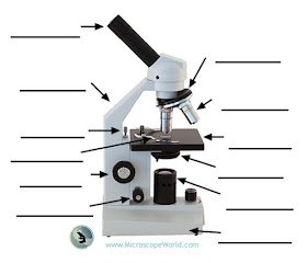
Labeling the Parts of the Microscope
Labeling the parts of the microscope is a common activity in schools. Below you will find both the label the microscope activity worksheet as well as one with answers.
Microscope World has PDF printable versions of both microscope activities shown below that you can print and use for free here.
Microscope World has PDF printable versions of both microscope activities shown below that you can print and use for free here.
 |
| Label the Parts of the Microscope |
 |
| Label the Parts of the Microscope Answers |
Tuesday, October 6, 2015
Diphtheria under the Microscope
Corynebacterium Diphtheriae is a rod-shaped bacteria belonging to the order of Actinomycetales, which are typically found in soil, but also have pathogenic members such as Streptomyces and mycobacteria. It is a gram-positive, aerobic, non-motile and toxic producing bacteria.
Corynebacterium Diphtheriae is best known for causing the disease Diphtheria in human beings, which results from the production of the Diphtheria toxin in conjunction with an infection by a bacteriophage, which provides it with a toxin-producing gene. Diphtheria has been studied heavily, because historically it was a very deadly disease, especially for children where mortality rates were eighty percent before vaccines and antitoxin were developed. Since rats and mice are naturally immune to the Diphtheria toxin, it has been difficult to study in a lab.
Diseases resembling Diphtheria were described as early as the 4th century BCE by Hippocrates. It was named in 1826 by French physician Pierre Bretonneau, who gave it the Greek name for leather hide. This was a very appropriate name since the disease creates a leathery layer that grows on tonsils, throat and nasal passages.
By 1892 the first antiserum was developed by Emil Von Behring and ready for commercial production. He was later awarded the Nobel Prize in medicine in 1891.
In 1923 Alexander Glenny, Barbara Hopkins and Gaston Ramon produced a vaccine for Diphtheria. They found that formaldehyde could prevent the Diphtheria toxin from being toxic, and the non-toxic form still retained its antigenic qualities. This vaccine led to a large decline in the number of cases of Diphtheria. In 2014 there was only one recorded case of Diphtheria in the United States.
The images below of Corynebacterium Diphtheriae were captured using a lab research microscope and a CCD microscopy camera.
 |
| Corynebacterium Diphtheriae under the microscope at 40x. |
 |
| Corynebacterium Diphtheriae under the microscope at 100x. |
 |
| Corynebacterium Diphtheriae under the microscope at 400x. |
 |
| Corynebacterium Diphtheriae under the microscope at 400x captured with a Plan Fluor Objective. |

